
Originally published September 4, 2024. Reviewed and updated August 10, 2026.
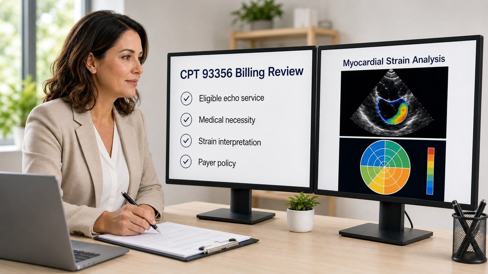
CPT® code 93356 is used to report myocardial strain imaging performed with speckle-tracking echocardiography. It is an add-on code, which means it cannot be reported by itself. The claim must include an eligible primary echocardiography service performed for the same patient by the same practitioner on the same date of service.
That coding rule is only the beginning. A valid parent code does not establish medical necessity, guarantee coverage, or guarantee payment. In 2026, practices also need to account for payer-specific coverage criteria, setting-specific payment rules, clear strain findings in the report, and Medicare's one-unit medically unlikely edit.
This guide summarizes the current federal coding and payment files, explains what a defensible report should contain, and highlights workflow considerations for practices evaluating strain-capable cardiovascular ultrasound systems.
What changed for CPT 93356 in 2026?
CPT 93356 remains an add-on service, but the federal payment and claim-edit files used to validate billing and estimate reimbursement have been refreshed. This guide now reflects:
- The January 2026 Medicare Physician Fee Schedule RVUs and separate conversion factors for qualifying Advanced APM participants and other clinicians.
- The July 2026 Medicare NCCI list of acceptable primary services and the practitioner medically unlikely edit of one unit.
- The July 2026 Hospital Outpatient Prospective Payment System status indicator N, under which the outpatient hospital service is packaged rather than separately paid.
- The 2025 ASE/EACVI clinical consensus statement, including updated guidance for acquisition consistency, serial comparison, cardio-oncology, and interpretation.
These updates do not create automatic coverage or guarantee payment. Practices still need to verify the patient's plan, medical-necessity criteria, authorization requirements, documentation, and the files effective on the date of service.
CPT 93356 at a glance
| Question | 2026 answer |
|---|---|
| What does it report? | Myocardial strain imaging performed with speckle tracking and interpreted as part of an eligible echocardiography service. |
| Can it be billed alone? | No. CMS classifies 93356 as a Type 1 add-on code with a defined list of acceptable primary services. |
| How many units? | The July 2026 CMS practitioner MUE is 1 unit per practitioner, patient, and date of service. |
| What is the national Medicare estimate? | Approximately $36.74 in a nonfacility setting and $10.02 in a facility setting for non-QP clinicians, before geographic adjustment. QP estimates are approximately $36.92 and $10.07. |
| Is the hospital outpatient technical service paid separately? | Under the July 2026 OPPS Addendum B, 93356 has status indicator N, meaning the hospital outpatient service is packaged rather than separately paid. |
| Does a fee-schedule amount guarantee payment? | No. Coverage, medical necessity, documentation, contract terms, claim edits, and prior-authorization rules still apply. |
What is CPT code 93356?
CPT 93356 identifies myocardial strain imaging that uses speckle tracking to quantify myocardial mechanics. In everyday echocardiography, the most familiar strain measurement is left ventricular global longitudinal strain, or LVGLS, although strain imaging can also evaluate regional mechanics and other chambers when clinically appropriate.
The “+” designation matters: 93356 is an add-on code. CMS explains that a Type 1 add-on code is eligible for payment only when one of its specifically listed primary codes is also eligible for payment to the same practitioner for the same patient on the same date of service. Never choose a parent code simply because it appears on the acceptable list; the parent code must accurately describe the echocardiography service that was actually performed and documented.
Which parent codes can be reported with 93356 in 2026?
The July 2026 CMS NCCI add-on-code file lists the following primary codes for 93356:
| Primary code | Practical description |
|---|---|
| 93303 | Complete transthoracic echocardiography for congenital cardiac anomalies |
| 93304 | Follow-up or repeat transthoracic echocardiography for congenital cardiac anomalies |
| 93306 | Complete transthoracic echocardiography including spectral and color Doppler |
| 93307 | Complete transthoracic echocardiography without spectral or color Doppler |
| 93308 | Follow-up or limited transthoracic echocardiography |
| 93350 | Stress echocardiography imaging, interpretation, and report |
| 93351 | Stress echocardiography including the associated supervision and monitoring described by the code |
| C8921, C8922, C8923, C8924, C8928, C8929, C8930 | Certain hospital outpatient echocardiography services, including contrast-enhanced variants |
This is broader than many older summaries, which omit 93307 and the eligible hospital outpatient C-codes. Because CMS can revise NCCI files quarterly, confirm the version applicable to the date of service rather than relying on a saved list.
How many units of 93356 can be reported?
The July 2026 CMS practitioner MUE for 93356 is 1. For most claims, report one unit per practitioner, patient, and date of service. An MUE is a claim-edit threshold, not a coverage rule or a blanket authorization to bill one unit whenever strain software is used.
2026 Medicare payment estimates for CPT 93356
CMS introduced separate 2026 Physician Fee Schedule conversion factors for qualifying Advanced APM participants (QPs) and other clinicians. In the January 2026 RVU file, 93356 has 1.10 total nonfacility RVUs and 0.30 total facility RVUs.
Multiplying those unadjusted national RVUs by the applicable conversion factor produces the following estimates:
| 2026 PFS category | Total RVUs | Unadjusted national estimate |
|---|---|---|
| Nonfacility, non-QP conversion factor | 1.10 | $36.74 |
| Nonfacility, QP conversion factor | 1.10 | $36.92 |
| Facility, non-QP conversion factor | 0.30 | $10.02 |
| Facility, QP conversion factor | 0.30 | $10.07 |
These are educational national estimates, not promised allowed amounts. CMS calculates locality-specific rates by applying geographic practice cost indices to the work, practice-expense, and malpractice components. Use the current CMS Physician Fee Schedule Look-Up Tool or the definitive files from your Medicare Administrative Contractor for the service location and date of service.
The facility amount above is the Physician Fee Schedule amount for the practitioner service in a facility setting. It is not a separate hospital technical-component payment. In the July 2026 Hospital Outpatient Prospective Payment System Addendum B, 93356 has status indicator N, so the hospital outpatient service is packaged into payment for the primary service rather than paid separately under OPPS.
Also remember that an allowed amount is not the same as net collected revenue. Coverage decisions, patient cost sharing, payer contracts, claim edits, write-offs, and collection performance all affect the final result. A responsible business forecast should start with verified payer mix and historical adjudication—not an assumption that a fixed percentage of echo patients qualifies for strain imaging.
Does Medicare or commercial insurance cover 93356?
A published RVU and payment amount do not create a universal coverage guarantee. Original Medicare claims still must satisfy medical-necessity, documentation, coding, and other applicable rules. Medicare Advantage and commercial plans may use their own medical policies, diagnosis requirements, prior-authorization rules, and network contracts.
Commercial policies can be quite narrow. As one current example, Aetna's adult policy identifies defined scenarios involving cardio-oncology, heart-transplant evaluation or surveillance, and left ventricular hypertrophy with both unclear etiology and concern for infiltrative cardiomyopathy. It considers other uses unproven or not medically necessary. That example should not be applied to a different payer or plan.
Before the study, confirm:
- Whether the patient's exact plan covers 93356 for the documented indication.
- Whether prior authorization or pre-notification is required.
- Which diagnosis codes the payer accepts for that indication.
- Whether the primary echocardiogram and the add-on service must be billed by the same entity or practitioner under the applicable rule.
- Whether the place of service changes separate-payment or packaging treatment.
- Whether the payer has frequency limits or its own edits in addition to CMS NCCI edits.
Clinical usefulness and payer coverage are related but separate questions. A professional-society consensus statement can support clinical rationale, but it does not override a payer's benefit language or medical policy.
CPT 93356 documentation requirements: what should the report include?
The medical record should make it easy for a reviewer to understand why strain imaging was needed, how it was performed, what it showed, and how the result affected the interpretation. A defensible workflow generally includes:
- Clinical indication and medical necessity. State the patient-specific question being evaluated. Avoid generic phrases such as “strain performed” without explaining why it was reasonable for this patient.
- Eligible primary study. Document the complete, limited, congenital, or stress echocardiography service that accurately reflects the work performed.
- Technique. Identify speckle-tracking myocardial strain imaging. Include the chamber and measurement approach when relevant.
- Quantitative findings. Report the strain value or values, such as LVGLS, and any regional pattern that is clinically relevant.
- Interpretation. Explain whether the findings are normal, abnormal, changed from baseline, limited by image quality, or otherwise clinically meaningful. A number without interpretation is weak documentation.
- Comparison. For serial studies, identify the prior examination and describe the absolute or relative change using a consistent method when possible.
- Technical limitations and consistency. Note suboptimal tracking or acquisition limitations. Recording the analysis platform and software version can improve reproducibility in longitudinal follow-up.
- Authenticated report. Ensure the interpreting clinician completes and signs the report in accordance with payer and facility requirements.
Do not let an automated measurement populate the final report without clinician review. Software availability alone does not establish that the separately reportable service was medically necessary, successfully performed, or interpreted.
A concise documentation example
Myocardial strain imaging was performed using two-dimensional speckle tracking because the patient is receiving potentially cardiotoxic therapy. LV global longitudinal strain was measured from standard apical views and compared with the baseline examination. The report documents the current value, relative change, image-quality limitations, and the interpreting clinician's conclusion.
Adapt any template to the actual patient, study, payer, and documentation rules. Do not copy a stock statement into records when the work was not performed.
What changed in the 2025 ASE/EACVI strain consensus statement?
The American Society of Echocardiography and European Association of Cardiovascular Imaging published an updated clinical consensus statement in November 2025. It addresses the use of strain echocardiography across heart failure, cardio-oncology, ischemic and valvular disease, cardiomyopathies, transplantation, congenital heart disease, and other clinical settings.
Several practical points are especially relevant to longitudinal echo programs:
- Normal limits vary with the population, vendor, analysis method, and software version. Avoid treating a single universal cutoff as appropriate for every patient and platform.
- Sequential comparison is often more useful than an isolated value. The consensus states that a relative LVGLS change of roughly 10% to 15% is likely to be significant when serial studies use a consistent approach.
- In cardio-oncology, a conservative relative LVGLS decrease of 15% during therapy is considered evidence of preclinical cardiac dysfunction in the clinical context described by the statement.
- Follow-up should use the same vendor's analysis software and, when feasible, the same software release because intervendor and interversion differences can affect values.
- Image quality, correct timing, and expert review remain important even when AI-assisted tools accelerate analysis.
These clinical recommendations strengthen the case for consistent acquisition and reporting. They do not automatically establish coverage for CPT 93356, which still depends on the patient's plan and the documented indication.
Common reasons 93356 claims are denied
Denials often trace to one of a few preventable gaps:
- 93356 was submitted without an eligible primary code, or the primary service was denied.
- The report contains a strain value but no patient-specific indication or interpretation.
- The diagnosis on the claim does not match the condition documented in the record or the payer's policy.
- Required prior authorization or notification was not obtained.
- More than one unit was submitted for the same practitioner, patient, and date of service.
- The practice expected a separate hospital outpatient technical payment even though OPPS packaging applies.
- The claim used rules, payment rates, or NCCI files from the wrong year or quarter.
What to do after a denial
- Read the remittance advice and identify whether the issue is coverage, coding, authorization, documentation, bundling, or a clerical error.
- Retrieve the policy and edit files that were effective on the date of service—not just the payer's current webpage.
- Confirm that the primary echocardiography claim was accepted and that the parent code accurately represents the documented study.
- Assemble the order, clinical notes, complete echo report, strain measurements and interpretation, authorization record, and any relevant policy language.
- Correct and resubmit a true claim error. Use the payer's formal appeal process when disputing a medical-necessity or coverage decision.
- Track denial reasons by payer and indication so scheduling, authorization, documentation, and coding workflows can be improved.
Peer-reviewed evidence and the 2025 ASE/EACVI statement may help explain clinical rationale, but an appeal should address the payer's stated reason and policy rather than relying on general evidence alone.
Strain-imaging workflow on GE Vivid systems
Reliable strain programs depend on good acquisition, consistent analysis, a documented clinical interpretation, and a workflow that staff can repeat. Equipment features can make that work faster and more consistent, but they do not determine coverage or guarantee reimbursement.
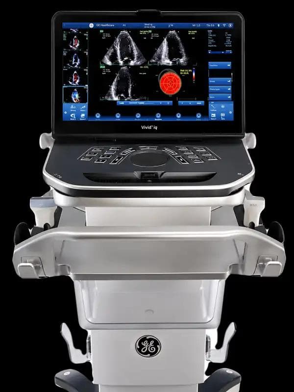
For practices that need a portable cardiovascular platform, the GE Vivid IQ portable cardiovascular ultrasound system supports GE's Easy AFI LV workflow and is suited to mobile scanning, satellite locations, and space-constrained environments.
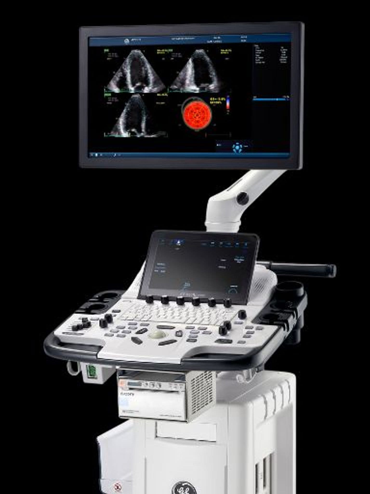
For practices that prefer a compact cart-based system, the GE Vivid T9 cardiac ultrasound system combines cardiovascular and shared-service capabilities and supports Easy AFI LV on applicable configurations.
Options and software packages can vary by model, configuration, and system generation. Confirm that the specific new, preowned, or rental system includes the strain applications, probes, software level, reporting integration, training, and service support your program requires.
Frequently asked questions about CPT 93356
Can CPT 93356 be billed by itself?
No. It is a Type 1 add-on code under Medicare NCCI rules and must be reported with an eligible primary echocardiography code for the same patient, practitioner, and date of service.
What are the valid 93356 parent codes in 2026?
The July 2026 CMS file lists CPT codes 93303, 93304, 93306, 93307, 93308, 93350, and 93351, plus HCPCS codes C8921, C8922, C8923, C8924, C8928, C8929, and C8930. Always use the file applicable to the date of service.
How many units of 93356 can Medicare practitioners report?
The July 2026 practitioner MUE is 1 unit per practitioner, patient, and date of service for the vast majority of correctly reported claims.
How much does Medicare pay for 93356 in 2026?
Using the January 2026 national RVUs and conversion factors, the unadjusted estimate is approximately $36.74 nonfacility and $10.02 facility for non-QP clinicians. QP estimates are approximately $36.92 and $10.07. Actual rates vary by locality and other payment rules.
Does Medicare's fee schedule mean 93356 is always covered?
No. A payment amount does not guarantee coverage. The service still must satisfy medical necessity, documentation, coding, and any applicable Medicare or payer requirements.
Is there a separate hospital outpatient payment for 93356?
Not under the July 2026 OPPS Addendum B. Status indicator N means the hospital outpatient service is packaged into payment for the primary service.
Is C9762 the MRI equivalent of 93356?
No. C9762 describes strain-encoded cardiac magnetic resonance imaging. CPT 93356 applies to myocardial strain imaging performed with echocardiographic speckle tracking. The modalities and coding pathways are different.
Does owning strain-capable software make every study billable?
No. The service must be clinically indicated, actually performed, interpreted, documented, paired with an eligible parent service, and covered under the patient's plan. Product capability does not establish medical necessity.
Authoritative sources
- CMS Medicare NCCI Add-on Code Edits
- CMS Medicare NCCI Medically Unlikely Edits
- CMS CY 2026 Physician Fee Schedule Final Rule fact sheet
- CMS January 2026 Relative Value Unit files
- CMS Physician Fee Schedule Look-Up Tool
- CMS July 2026 OPPS Addendum B
- 2025 ASE/EACVI Clinical Applications of Strain Echocardiography consensus statement
- Aetna myocardial strain imaging policy