
GE Ultrasound Feature
3D Ultrasound
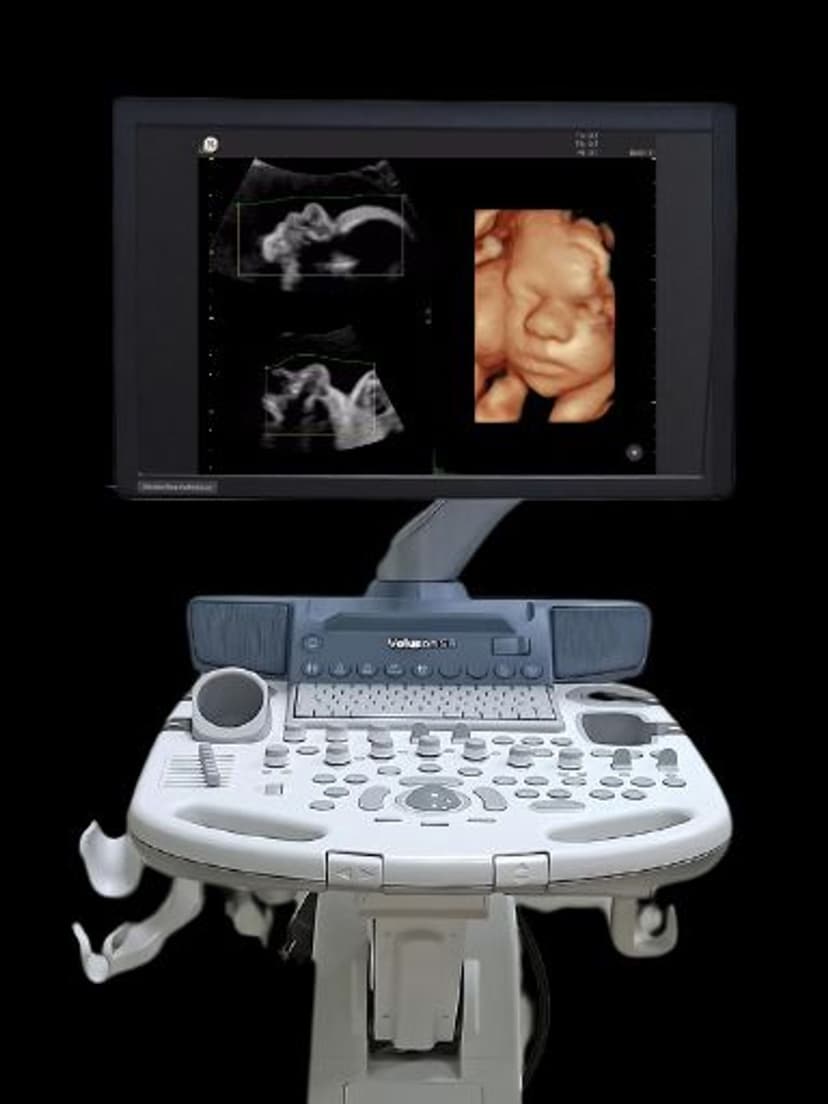
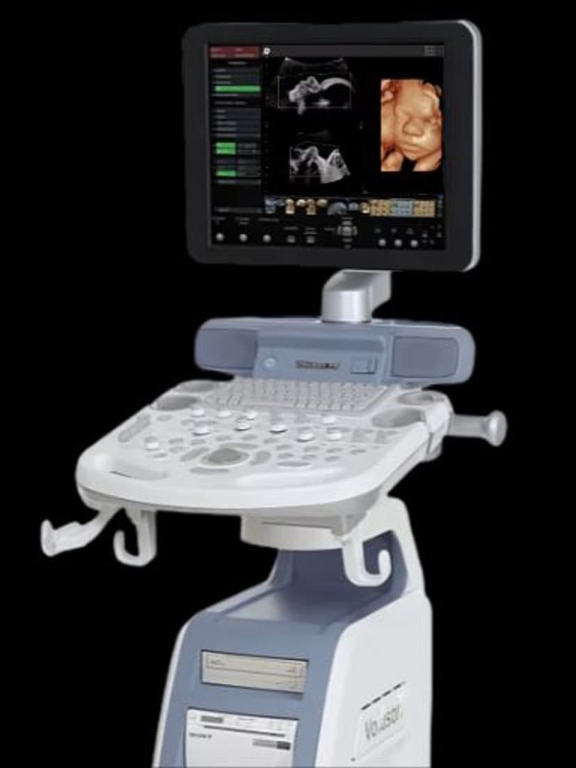
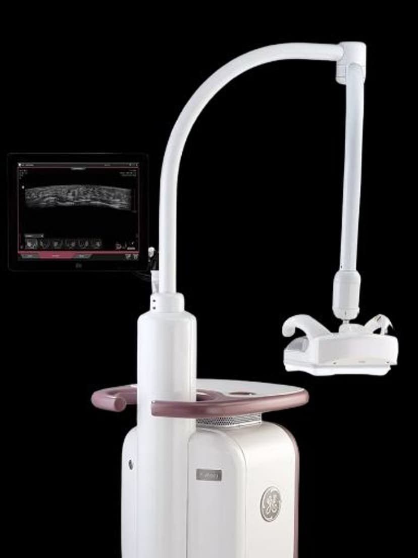
Imaging Technology3D Ultrasound on GE systems captures echo data from multiple angles and reconstructs a volumetric image that clinicians can view, rotate, and slice in any plane. On Voluson-series systems, 3D acquisition pairs with HDlive rendering to produce photorealistic surface images with natural depth and lighting. On the Invenia ABUS, 3D volume scanning provides automated whole-breast imaging for supplemental screening. Clinicians examine anatomy in coronal, sagittal, and axial planes from a single acquisition, including oblique and off-axis views impossible to obtain in standard 2D imaging.

Key Benefits
Why 3D Ultrasound matters
Multiplanar anatomy from a single acquisition
One 3D volume dataset provides coronal, sagittal, axial, and oblique views without rescanning. The coronal plane, inaccessible in standard 2D imaging, becomes available for assessing structures like the uterine cavity, fetal brain, and renal anatomy.
HDlive surface rendering for fetal anatomical detail
GE's HDlive technology applies realistic lighting and depth to 3D surface images. In obstetric imaging, this produces detailed views of fetal facial features, extremities, and surface anatomy that support detection of structural abnormalities and improve parent communication.
Automated whole-breast volume scanning on Invenia ABUS
On the Invenia ABUS system, 3D volume acquisition captures the entire breast in a standardized scan. Radiologists review the volume slice by slice, improving detection of small lesions in dense breast tissue where mammography has reduced sensitivity.
Offline review and remote consultation capability
Stored 3D volume datasets can be reviewed, resliced, and measured after the patient leaves. This supports remote reading workflows where a specialist reviews volumes acquired at a satellite clinic, and enables second-opinion consultations without requiring the patient to return for a repeat exam.
About 3D Ultrasound
3D Ultrasound technology acquires a volume of echo data by sweeping the ultrasound beam through the target anatomy. This can be performed using dedicated volume probes that sweep mechanically, or through freehand acquisition where the system registers sequential 2D frames based on probe motion. The captured volume is reconstructed into a three-dimensional dataset that clinicians navigate using multiplanar reformatting. Any arbitrary plane through the volume can be displayed, including the coronal plane which is not directly accessible in conventional 2D ultrasound. In obstetric imaging, 3D acquisition with HDlive rendering produces surface-rendered images of fetal anatomy used for detecting cleft lip, assessing limb morphology, and evaluating complex structural abnormalities. Multiplanar views of the fetal brain, spine, and heart reveal anatomy not visible in standard 2D sweeps. On the Invenia ABUS system, 3D volume acquisition takes a different approach: an automated breast scanner captures a full volume of breast tissue in a standardized, reproducible scan. This application targets supplemental breast cancer screening in women with dense breast tissue, where mammographic sensitivity is reduced. The 3D volume data can be reviewed slice by slice through the entire breast, improving detection of small masses that may be obscured in dense parenchyma.
Our Partners


Want 3D Ultrasound in your practice?
Request a quote for a system that includes this feature.