
GE Ultrasound Feature
4D Imaging
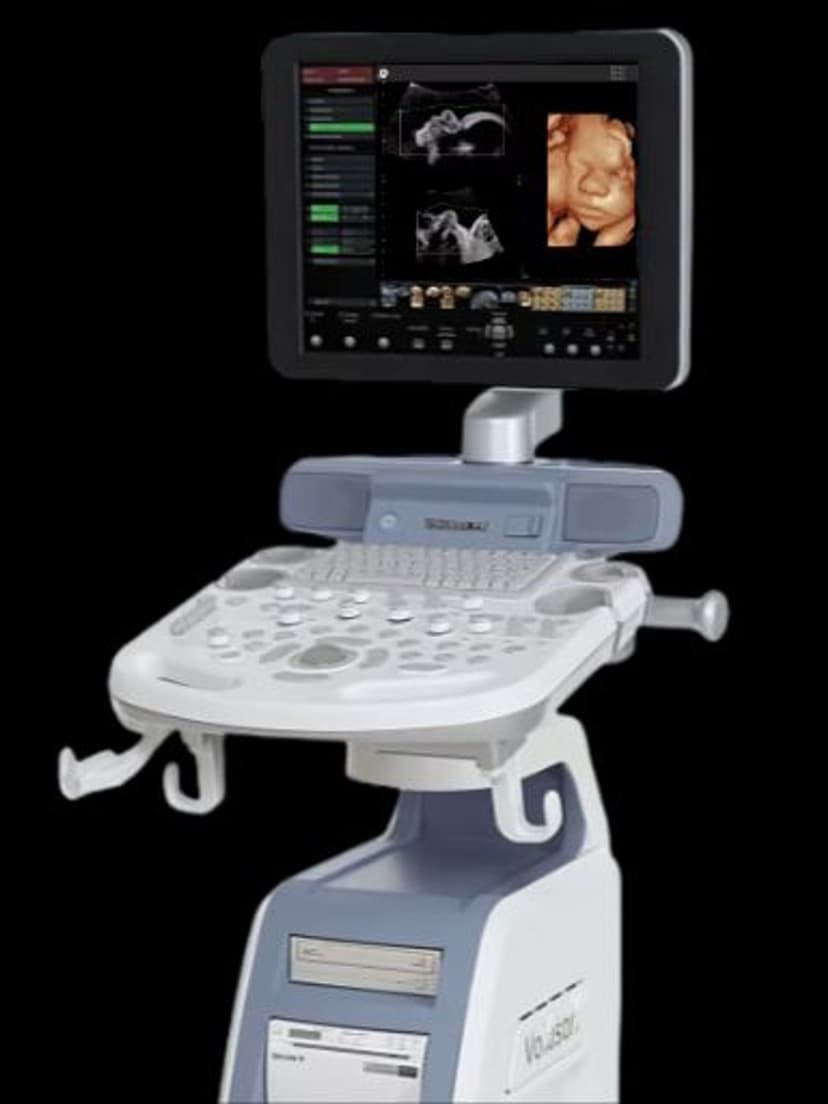
Imaging Technology4D Imaging on GE ultrasound systems captures continuous 3D volume data and renders it in real time, producing live-motion volumetric images. The technology acquires a series of 3D frames in rapid succession, creating a moving picture that shows anatomical structures as they function. In obstetrics, 4D Imaging visualizes fetal movements, facial expressions, and limb activity in real time. In cardiology, it captures cardiac wall motion and valve dynamics across the full cardiac cycle. On GE Vivid and Voluson systems, 4D Imaging works with HDlive rendering to produce photorealistic volume images with natural-looking depth and lighting effects.

Key Benefits
Why 4D Imaging matters
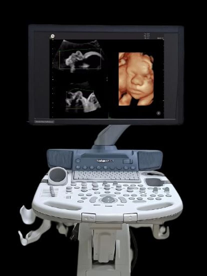
Live-motion fetal assessment beyond static 2D views
4D Imaging shows fetal movements, facial expressions, and limb activity in real time. Clinicians observe swallowing, breathing, and extremity motion patterns not visible in standard 2D imaging, aiding detection of structural and functional abnormalities.
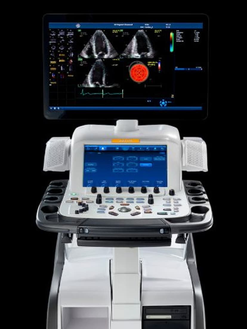
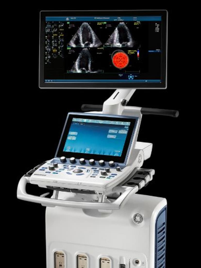
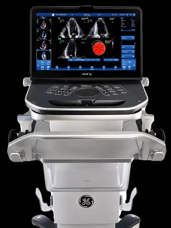
Dynamic cardiac wall and valve visualization
In echocardiography, 4D Imaging captures the full cardiac cycle in a volumetric dataset. Clinicians assess wall motion abnormalities, valve morphology, and chamber geometry in real time without the mental reconstruction required when interpreting multiple 2D planes.
Stronger patient engagement and communication
The photorealistic, live-motion images are immediately understandable to patients. In obstetric settings, parents see their baby moving in real time, which improves patient satisfaction and strengthens the clinical relationship between provider and family.
Reduced need for repeat exams in difficult fetal positions
When fetal position limits 2D assessment, 4D volume data often captures the anatomy from an accessible angle. Clinicians can rotate and review the 4D dataset to find diagnostic views without scheduling a follow-up scan to wait for fetal repositioning.
About 4D Imaging
4D Imaging extends 3D ultrasound by adding temporal resolution. Where 3D captures a single static volume at one moment, 4D acquires volumes continuously at frame rates sufficient to display smooth real-time motion. This requires volume probes capable of rapid mechanical or electronic scanning, paired with processing power to reconstruct and render each volume frame without noticeable lag. On GE systems, 4D Imaging uses dedicated volume transducers that sweep through the anatomy automatically, eliminating the manual freehand sweep technique required for static 3D. The resulting live video shows structures in motion: fetal limbs moving, heart valves opening and closing, or diaphragms contracting during respiration. In obstetric practice, 4D Imaging improves the assessment of fetal anatomy by revealing surface features and movement patterns not visible in 2D imaging alone. Clinicians use it to evaluate fetal facial anatomy, detect cleft lip, assess limb morphology, and observe swallowing or breathing movements. In cardiac imaging, 4D echocardiography provides volumetric assessment of chamber function and valve dynamics, supporting quantitative measurements like real-time ventricular volumes. GE pairs 4D acquisition with HDlive rendering on Voluson systems and with volume-based measurement tools on Vivid systems.
Our Partners


Want 4D Imaging in your practice?
Request a quote for a system that includes this feature.