
GE Ultrasound Feature
Adv. Cardiac Imaging
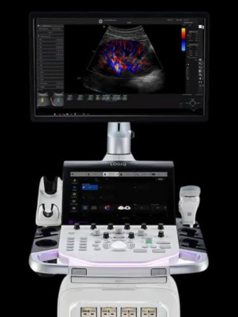
CardiacAdvanced Cardiac Imaging is a capability tier that brings echocardiography-grade visualization to the LOGIQ Totus general imaging console. It combines high-frequency Doppler, multi-plane views, and myocardial motion analysis to capture structural and functional assessments of the heart chambers, valves, and great vessels in real time. Practices that scan a mix of abdominal, vascular, and cardiac patients can cover echo workflows on a single general-imaging platform, supporting differential diagnosis in conditions like valve disease, heart failure, and suspected cardiomyopathy without routing patients to a dedicated cardiovascular suite.

Key Benefits
Why Adv. Cardiac Imaging matters
Turns an urgent echo request into a same-day read
Hospitals and outpatient practices can handle unplanned cardiac workups without scheduling around a dedicated echo lab's availability. Bedside and inpatient cardiac assessments stay inside the radiology department's existing rotation rather than being pushed to cardiology.
Flows echo measurements into DICOM-standard worksheets
Quantitative outputs populate the same structured reporting fields that reading physicians use for dedicated echo studies. Referring cardiologists get reports in a familiar format rather than a free-text interpretation from a general-imaging console.
Lets sonographers cross-cover cardiac without a second platform
One button layout and one measurement package cover abdominal, vascular, and cardiac exams on the same shift. Technologists trained on LOGIQ don't have to learn Vivid-specific controls to take a cardiac case.
Quantifies valve disease severity without a downstream referral
Pressure gradients, regurgitant jets, and wall motion scoring give the ordering physician enough data to triage a new murmur or suspected heart failure on the first encounter. Fewer patients leave with a pending-test note and come back for a separate appointment.
About Adv. Cardiac Imaging
On a LOGIQ Totus, Advanced Cardiac Imaging supports the same Doppler modes and measurement packages a cardiologist would expect: color flow, pulsed and continuous-wave Doppler across valves, M-mode for timing cardiac cycles, and Tissue Doppler for quantifying myocardial velocity. The combination produces a set of numbers — peak gradients across stenotic valves, regurgitant volumes, ejection fraction approximations, wall motion scores — that feed structured reporting worksheets. The practical significance for a general-imaging department is coverage: the same console that handled the morning's abdominal and OB list can take an urgent echo request in the afternoon without an equipment swap. For practices without a dedicated Vivid system, this extends the clinical range of their existing fleet rather than adding a second capital purchase.
Availability
Available on these systems
Our Partners


Want Adv. Cardiac Imaging in your practice?
Request a quote for a system that includes this feature.