
GE Ultrasound Feature
AI Auto Measure
CardiacAI Auto Measure uses machine learning to detect anatomical landmarks and automatically place measurement calipers on 2D and Doppler images during echocardiography exams. Instead of manually positioning calipers on chamber dimensions, wall thicknesses, and Doppler waveforms, the system identifies the correct measurement points and calculates values in a single step. This cuts the time spent on routine cardiac measurements and reduces the variability that comes with different operators placing calipers at slightly different locations. Cardiologists, sonographers, and POCUS users benefit from faster reporting and more reproducible results across studies.

Key Benefits
Why AI Auto Measure matters
Automated caliper placement across 2D and Doppler modes
AI Auto Measure detects measurement landmarks in both 2D and spectral Doppler images. It handles chamber dimensions, wall thicknesses, and flow velocities in a single workflow, covering the most time-consuming portion of a standard echo exam.
Reduced inter-operator measurement variability
Manual caliper placement varies from sonographer to sonographer, especially for measurements like LV internal diameter where millimeter differences affect clinical decisions. AI-driven placement applies the same landmark detection consistently, improving study-to-study comparability.
Faster exam completion for high-volume labs
Automating the measurement step in each standard view removes the cursor-position-click-repeat cycle that adds minutes to every study. Labs running 15+ daily echos see real throughput gains without sacrificing measurement quality.
Auto-populated structured reports
Measurement values transfer directly into the reporting workflow as they are captured, eliminating the manual data entry step at the end of the exam. Sonographers finish with a near-complete report by the time the last view is acquired, reducing post-exam documentation time.
About AI Auto Measure
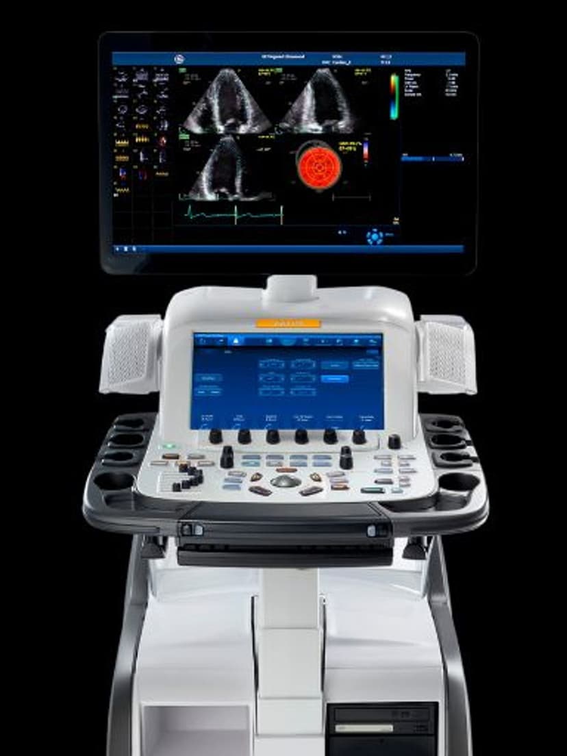
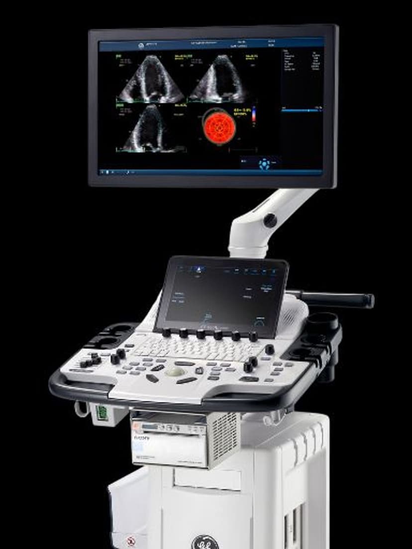
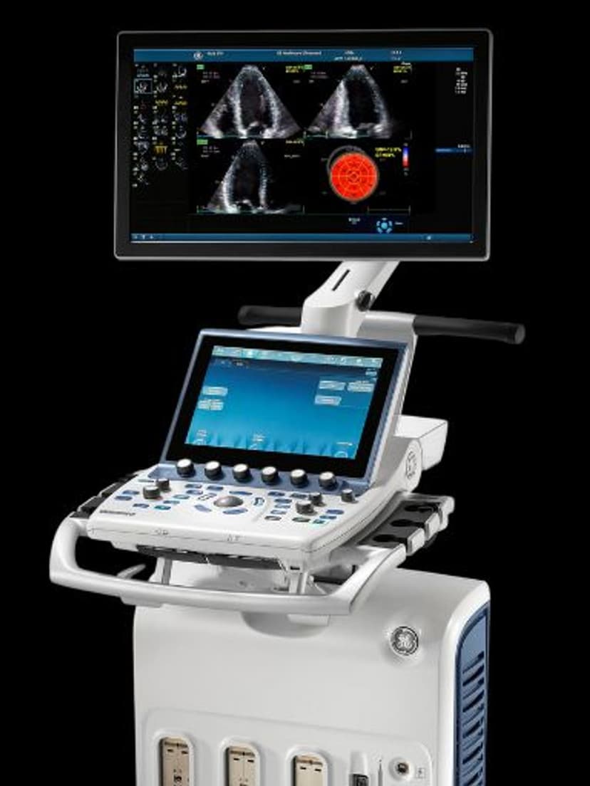
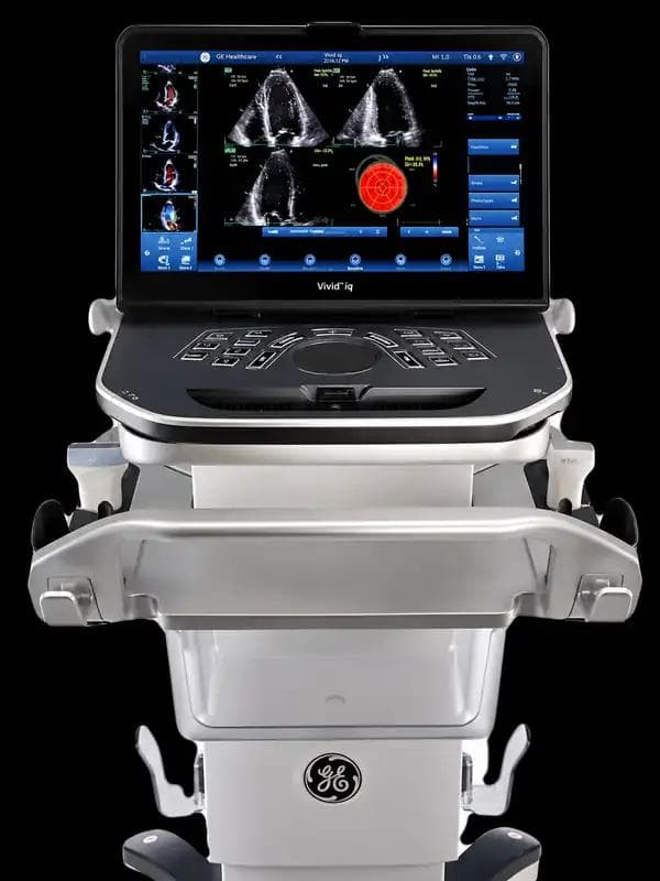
AI Auto Measure applies deep learning models trained on echocardiographic datasets to recognize standard cardiac views and their associated measurement landmarks. In 2D mode, the system identifies structures such as the interventricular septum, left ventricular internal diameter, and posterior wall thickness in parasternal long-axis views, and places calipers at end-diastole and end-systole. In Doppler mode, it traces mitral inflow E and A wave velocities, deceleration time, and LVOT outflow profiles. The operator reviews the AI-placed measurements, accepts or adjusts them, and moves to the next view. This workflow preserves clinical oversight while removing the repetitive cursor positioning that consumes a large share of echo exam time. The measurement values feed directly into the reporting package, so the structured report populates as the exam progresses. For high-volume echo labs processing 15 or more studies per day, the cumulative time savings per sonographer can reach 30 minutes or more daily.
Availability
Available on these systems
Our Partners


Want AI Auto Measure in your practice?
Request a quote for a system that includes this feature.