
GE Ultrasound Feature
AI-Based Tools
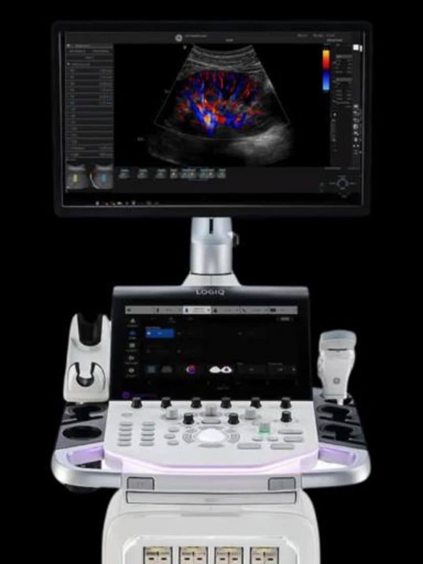
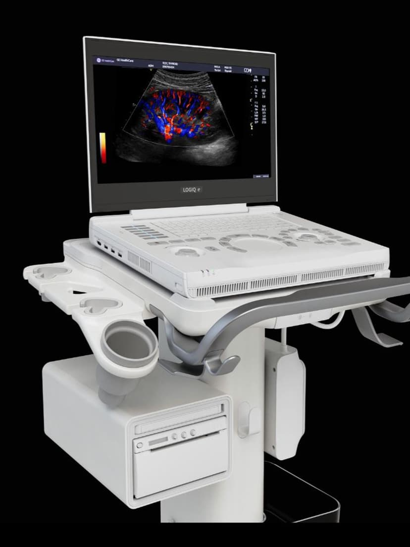
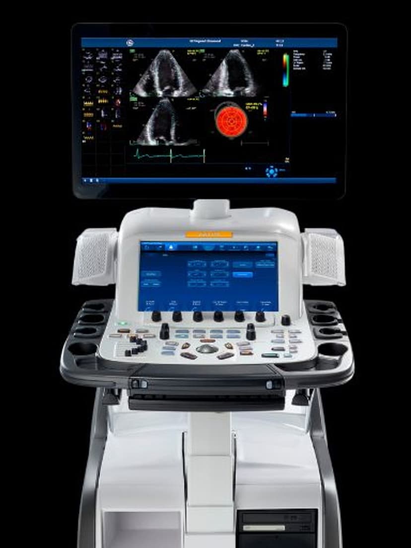
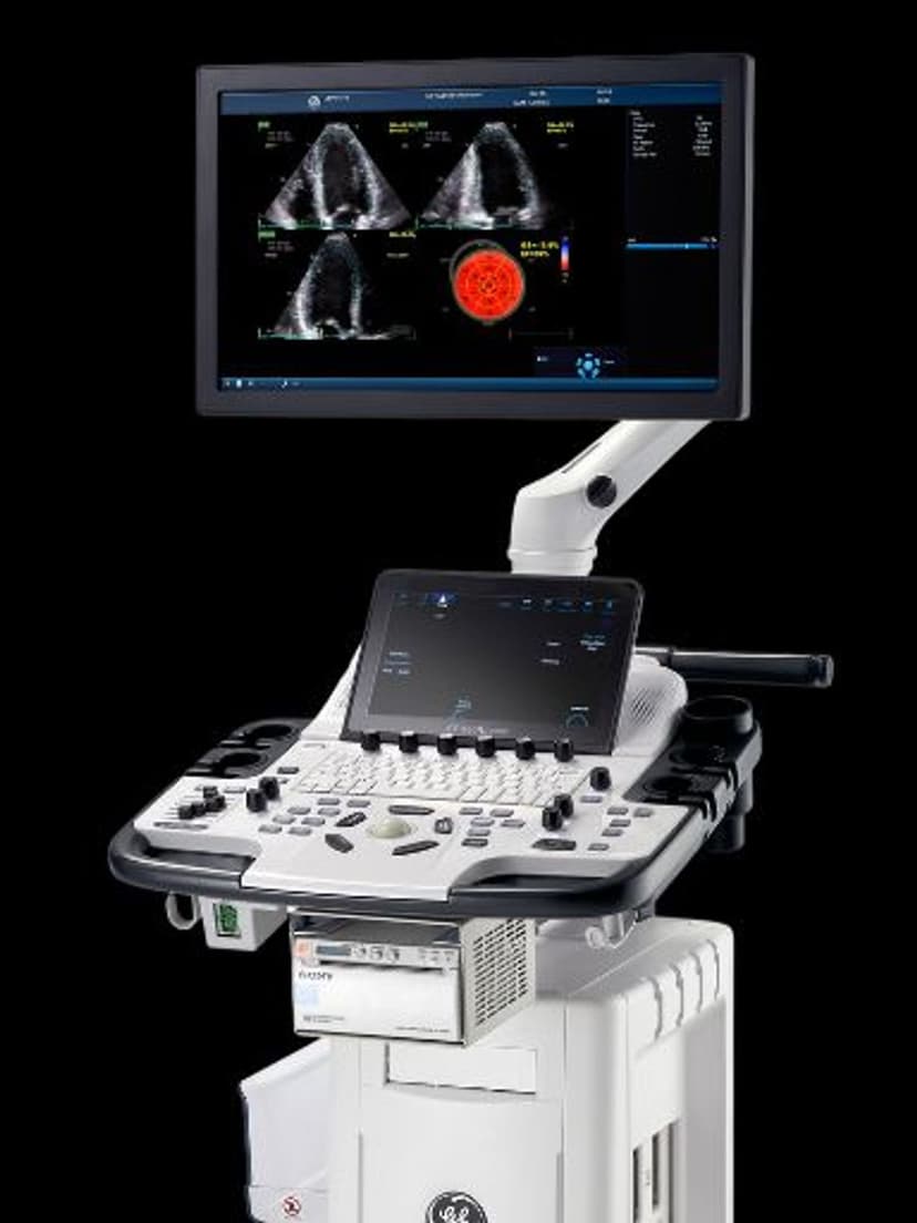
AI & AutomationAI-Based Tools is GE HealthCare's designation for the machine learning features built into its ultrasound systems, including automated measurements, image optimization, and workflow guidance that run during live scanning. The tools identify anatomical landmarks, place calipers, and adjust imaging parameters without manual input, reducing inter-operator variability and removing a common source of exam delay. Emergency departments, radiology reading rooms, and other high-volume settings rely on these features to hold measurement consistency and image quality steady across operators and shifts.

Key Benefits
Why AI-Based Tools matters
Cuts repetitive keystrokes on every exam
Auto-measurement and auto-caliper placement remove the dozens of manual button presses each study would otherwise require. Over a 15-to-25 patient day, that reduces both exam duration and the repetitive motion load on a sonographer's wrist and forearm.
Shortens the training curve for new operators
New sonographers and cross-trained physicians reach diagnostic-quality scans earlier because the system handles the parameter tuning and landmark recognition that traditionally takes months to internalize. Practices can put junior staff on independent scanning rotations sooner.
Catches missed views before the patient leaves
Protocol-aware workflow guidance flags incomplete studies in real time, not on QA review the next day. Fewer patient recalls, fewer reports held up waiting on repeat scans, and cleaner billing on the first pass.
Keeps multi-room fleets producing interchangeable studies
Practices running several ultrasound rooms get consistent measurement definitions and image presets regardless of which machine a patient is scanned on. Downstream readers, longitudinal comparisons, and prior-study matching all stay aligned without manual reconciliation.
About AI-Based Tools
GE's AI tools are trained on large image libraries to recognize standard views and anatomical structures during a live scan. When the probe reaches a target view, algorithms auto-detect landmarks, place calipers, and capture measurements — operations a sonographer would otherwise perform key-by-key. Image optimization runs in parallel: the system evaluates gain, depth, focal position, and TGC curves frame by frame and adjusts them to maintain consistent contrast and resolution as anatomy shifts. Workflow guidance prompts the operator through protocol steps, flags missing views, and carries settings forward between exams. Compared with manual optimization and post-hoc measurement, the approach removes the largest source of inter-operator variability: individual scanning technique. Consistency matters most where operators rotate frequently, because a reading radiologist comparing studies across shifts values reproducibility as much as peak image quality on any single scan. Clinicians can override any AI suggestion through customizable settings.


Our Partners


Want AI-Based Tools in your practice?
Request a quote for a system that includes this feature.