
GE Ultrasound Feature
AI-Driven Tools
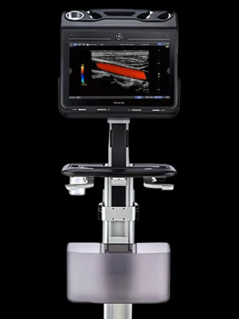
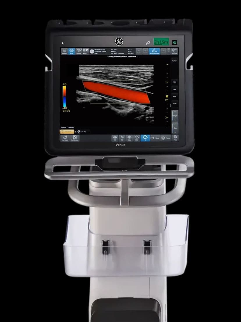
AI & AutomationAI-Driven Tools on the GE Venue Go is a collection of artificial intelligence algorithms that automate several steps of the point-of-care ultrasound workflow. These tools detect anatomical structures in real time, suggest and place measurements automatically, optimize imaging parameters based on the exam type, and annotate landmarks without manual input. For emergency physicians, hospitalists, and intensivists who need diagnostic answers fast, AI-Driven Tools reduce the number of manual adjustments between probe-on-patient and clinical decision.

Key Benefits
Why AI-Driven Tools matters
Automatic image optimization during live scanning
AI algorithms adjust gain, depth, and focus based on detected anatomy, so the image improves as the clinician scans. This removes much of the manual knob adjustment that less experienced operators struggle with at the point of care.
Measurements placed without manual caliper positioning
The system detects anatomical landmarks and positions measurement calipers automatically. This speeds up quantitative assessments and reduces inter-operator measurement variability in environments with rotating staff.
Real-time anatomical annotation
As the AI identifies organs, chambers, and vessels, it labels them on the display. This annotation supports faster interpretation during bedside assessments and creates structured documentation for the patient record.
Closes the skill gap for intermittent POCUS users
Emergency physicians, hospitalists, and intensivists who scan a few times per shift benefit most from AI assistance. The automated optimization and measurement tools help these clinicians achieve diagnostic-quality exams without daily scanning practice.
About AI-Driven Tools
AI-Driven Tools on the Venue Go represent GE's approach to making point-of-care ultrasound accessible to clinicians who scan intermittently rather than all day. The AI algorithms work in the background during live scanning: they identify anatomical structures as they appear in the field of view, apply appropriate imaging presets, and place measurement calipers on detected landmarks. For cardiac assessments, this can include automated chamber identification and ejection fraction estimation. For abdominal scans, the AI can detect and label organ boundaries. The tools also optimize gain, depth, and focus settings in real time based on what the system detects in the image, reducing the knob-turning that traditionally requires experience to get right. This automation layer is particularly valuable in settings where the operator may be an emergency physician or hospitalist who performs ultrasound as one of many clinical skills rather than a dedicated sonographer. By handling the technical adjustments, AI-Driven Tools let the clinician focus on the clinical question rather than image optimization.
Availability
Available on these systems
Our Partners


Want AI-Driven Tools in your practice?
Request a quote for a system that includes this feature.