
GE Ultrasound Feature
Augment Imaging
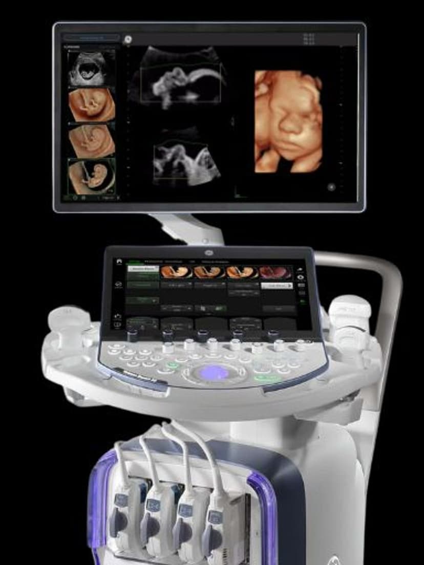
Imaging TechnologyAugment Imaging combines noise reduction algorithms with optimized beamforming to improve image clarity at depth. Available on the GE Voluson Expert, this technology reduces background clutter and improves tissue contrast, making it easier to visualize deep structures in patients with higher BMI or in late-term pregnancies where penetration is limited. The result is cleaner images with better tissue differentiation, reducing the need for repeat scans due to poor image quality.

Key Benefits
Why Augment Imaging matters
Clearer images at greater scanning depths
Optimized beamforming maintains lateral resolution and tissue contrast as depth increases. This is critical for late-term OB exams and patients with higher BMI where conventional imaging loses definition.
Reduced background noise without detail loss
The noise reduction algorithms selectively suppress clutter while preserving tissue boundaries and subtle anatomical details. This improves diagnostic confidence without the image smoothing that generic filters produce.
Fewer non-diagnostic exams in challenging patients
Better penetration and clarity mean fewer studies where image quality falls below what is needed for confident diagnosis. This reduces the need for repeat appointments or referrals to higher-end imaging.
Improved fetal anomaly screening accuracy
Clearer visualization of fetal cardiac structures, spine, and extremities during anatomy scans improves the detection rate for congenital abnormalities. Better images lead to earlier identification and more informed prenatal counseling.
About Augment Imaging
Ultrasound image quality degrades as imaging depth increases because the signal weakens and noise accumulates. This is a common challenge in OB/GYN imaging, particularly in the second and third trimesters when the fetus is further from the probe, and in patients with higher body mass index where tissue attenuation is greater. Augment Imaging addresses this through two mechanisms. First, noise reduction algorithms identify and suppress background clutter without removing clinically relevant tissue signals. Second, optimized beamforming focuses the transmit and receive beams to maintain lateral resolution at greater depths, preventing the image softening that typically occurs as depth increases. Together, these produce images with higher contrast-to-noise ratio and better definition of tissue boundaries. In practice, this means clearer views of fetal anatomy, improved placental assessment, and more confident evaluation of pelvic structures. The technology is particularly valuable during anatomy scans at 18-22 weeks, where clear visualization of fetal cardiac structures, spine, and extremities is essential for anomaly screening.
Availability
Available on these systems
Our Partners


Want Augment Imaging in your practice?
Request a quote for a system that includes this feature.