
GE Ultrasound Feature
Auto Contour
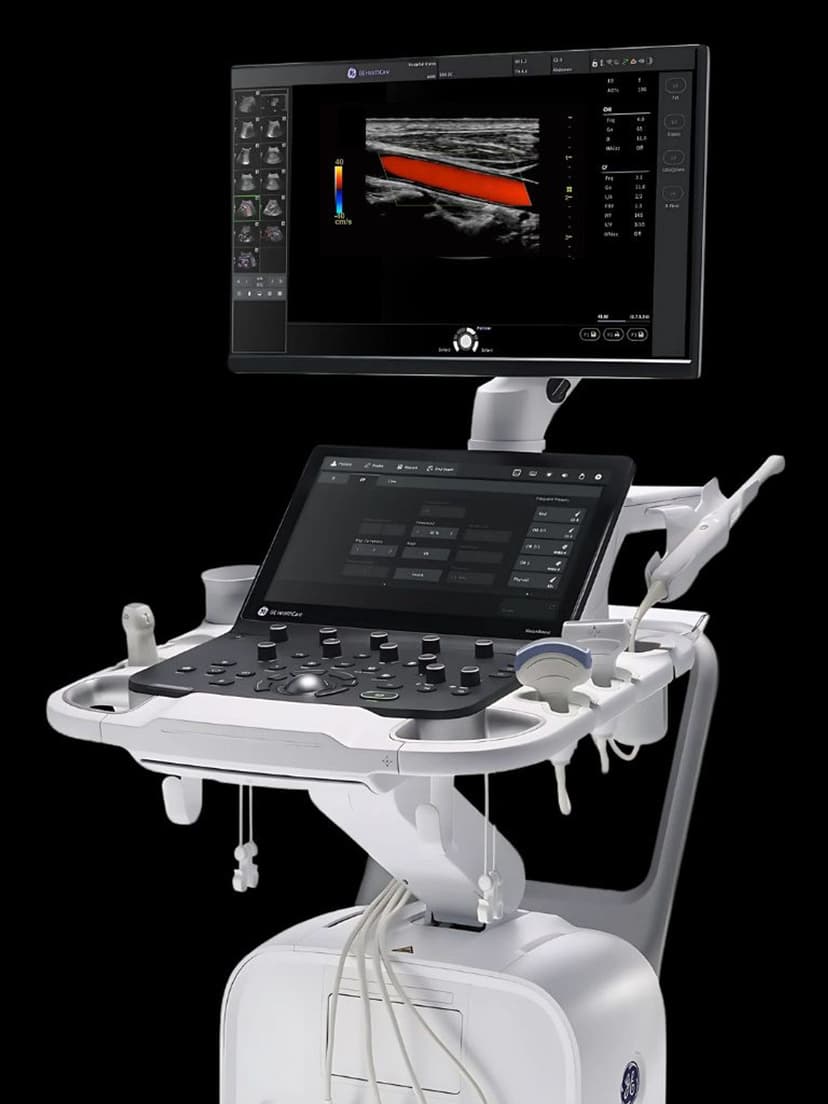
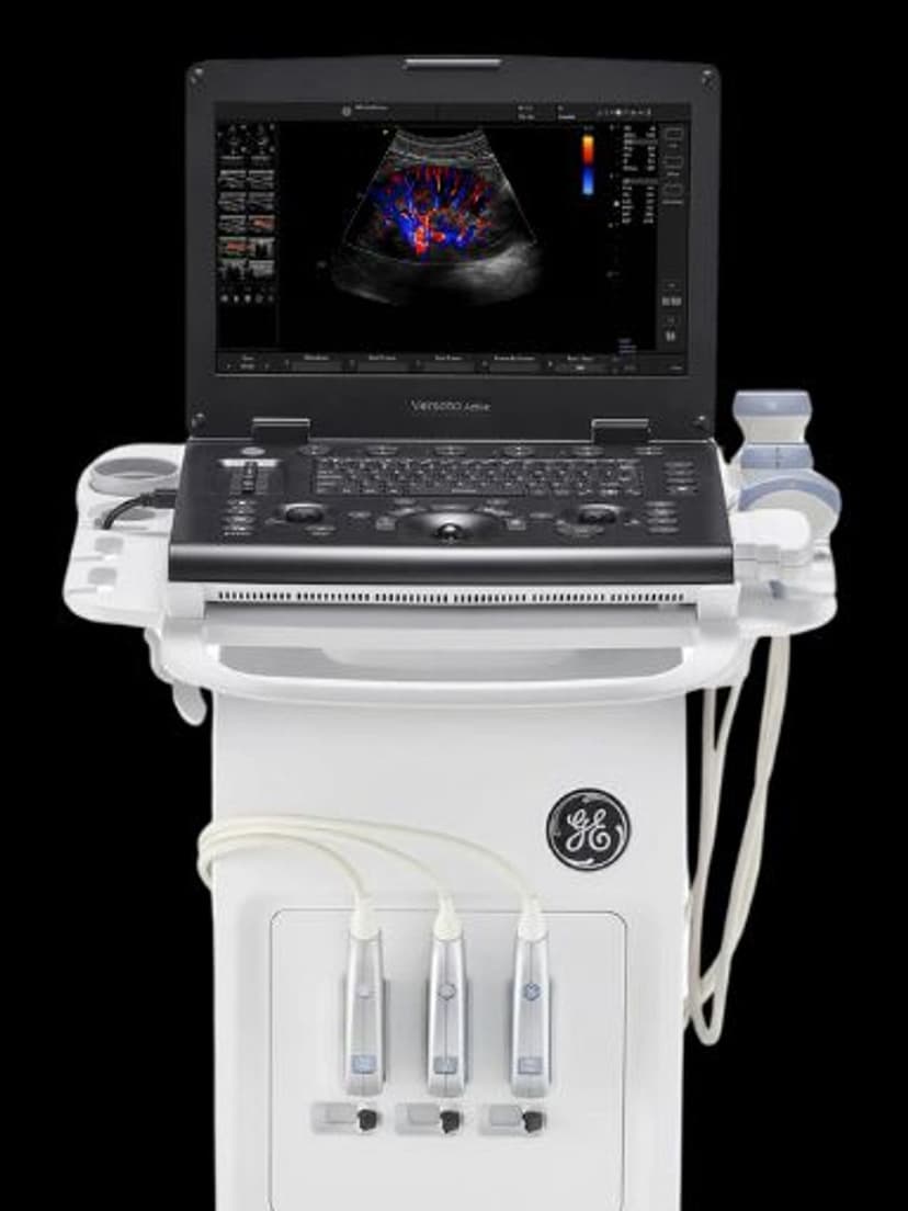
AI & AutomationAuto Contour uses AI algorithms to automatically detect and trace the boundaries of structures such as thyroid nodules, liver lesions, and other focal findings during ultrasound exams. Once the clinician identifies a structure of interest, Auto Contour places a precise outline around it and calculates dimensions, area, and volume measurements. This replaces manual caliper placement, which is time-consuming and varies between operators. Available on GE Versana systems, Auto Contour is particularly valuable in practices that perform high volumes of thyroid, breast, or abdominal imaging where consistent nodule measurements are needed for follow-up comparisons and reporting.

Key Benefits
Why Auto Contour matters
Automatic boundary detection replaces manual calipers
Auto Contour identifies and traces nodule margins based on echogenicity contrast with surrounding tissue. The algorithm places a contour around the entire structure rather than relying on two-point caliper measurements, capturing the actual shape and producing more accurate size estimates.
Consistent measurements for reliable follow-up comparisons
When tracking nodule size over serial exams, measurement consistency matters more than absolute accuracy. Auto Contour applies the same algorithmic approach each time, so size changes between studies reflect true growth rather than variations in how different operators place calipers.
Faster nodule documentation in multi-nodule exams
A thyroid exam with six or more nodules can require dozens of individual caliper placements. Auto Contour reduces per-nodule measurement time by automating boundary detection and dimension calculation, freeing the sonographer to focus on characterizing each finding rather than measuring it.
Supports standardized reporting criteria
Auto Contour measurements align with ACR TI-RADS size thresholds for thyroid nodules and similar standardized criteria for other organs. Consistent, reproducible measurements support structured reporting workflows and reduce the need for repeat measurements when values fall near decision thresholds.
About Auto Contour
Manual nodule measurement requires the sonographer to place two or three sets of calipers on each finding, measuring length, width, and anteroposterior diameter. For a thyroid exam with multiple bilateral nodules, this process can consume several minutes per patient and introduces variability depending on caliper placement technique. Auto Contour replaces this workflow by analyzing the B-mode image, identifying the boundary between the nodule and surrounding parenchyma based on echogenicity differences, and tracing the contour automatically. The system then calculates linear dimensions, cross-sectional area, and estimated volume from the contour data. Measurements follow ACR TI-RADS and other standardized size criteria, supporting consistent reporting across exams. In follow-up studies, Auto Contour's consistent measurement methodology makes it easier to detect true size changes versus measurement technique variation. The feature supports both 2D measurements on individual image frames and 3D volumetric measurements when volume data is available. For practices tracking nodule growth over time, the reduced inter-operator variability means that size changes between exams are more likely to reflect actual growth rather than differences in how two operators placed their calipers.
Availability
Available on these systems
Our Partners


Want Auto Contour in your practice?
Request a quote for a system that includes this feature.