
GE Ultrasound Feature
Auto EF 3.0
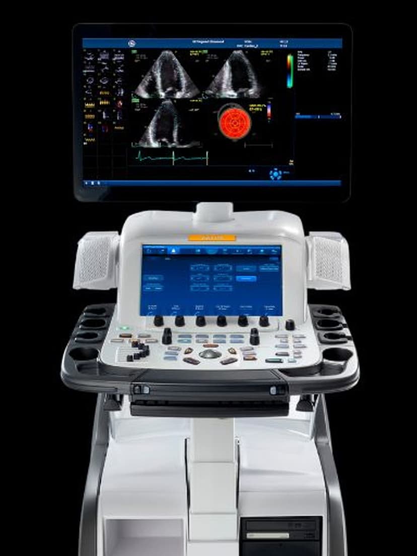
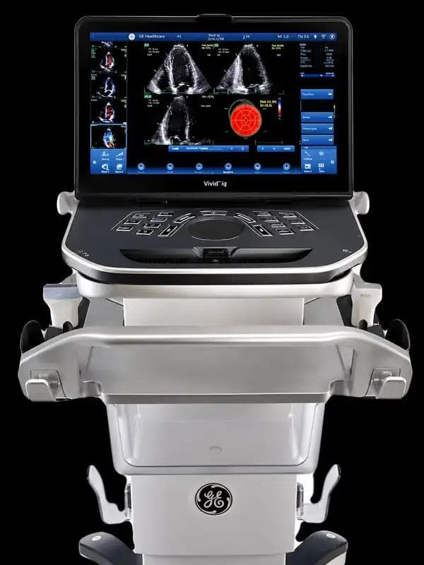
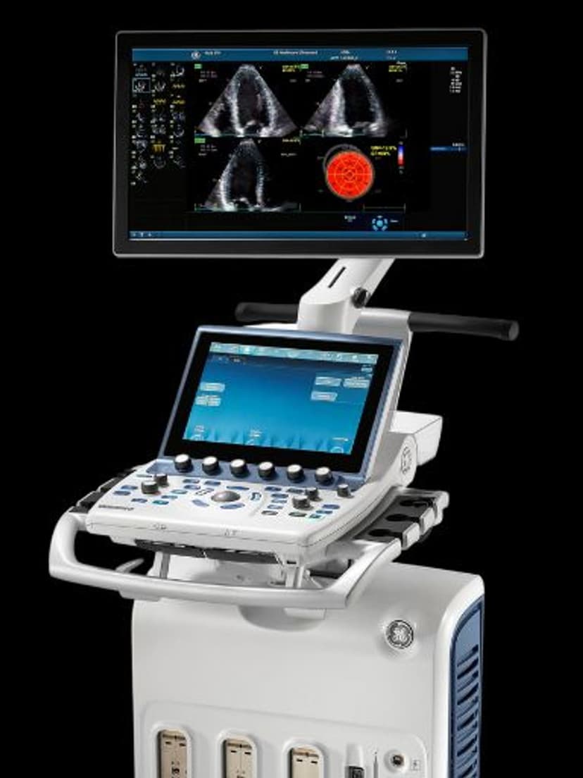
CardiacAuto EF 3.0 uses deep learning to automatically detect the left ventricular endocardial border, identify end-diastolic and end-systolic frames, and calculate ejection fraction using the biplane Simpson's method from apical two-chamber and four-chamber views. The entire measurement completes with a single button press in under 5 seconds. No ECG signal is required, making the tool usable in settings where ECG leads are impractical. Auto EF 3.0 produces consistent LVEF values across operators, addressing one of the biggest challenges in echocardiography: the 5-10% inter-observer variability that complicates serial patient monitoring and treatment decisions.

Key Benefits
Why Auto EF 3.0 matters
Guideline-compliant LVEF in under 5 seconds
One button press triggers automated frame selection, border tracing, and biplane volume calculation. The AI delivers a complete Simpson's method EF that follows ASE/EACVI quantification guidelines, replacing a multi-step process that takes 1-3 minutes manually.
Reduced inter-observer variability for serial monitoring
AI-driven border detection produces reproducible EF values regardless of which sonographer performs the study. This consistency is critical when tracking patients with heart failure, monitoring chemotherapy cardiotoxicity, or evaluating device therapy eligibility where small EF changes guide clinical decisions.
No ECG signal required
Auto EF 3.0 identifies cardiac cycle phases from the image data alone, eliminating the need for ECG leads. This makes the tool practical in point-of-care settings, during stress echo protocols, and in patients where ECG placement is difficult or impractical.
Faster onboarding for new cardiac sonographers
New staff produce expert-level EF measurements from day one. The AI handles the technically demanding steps of frame selection and border tracing, allowing less experienced sonographers to focus on image acquisition rather than manual quantification technique.
About Auto EF 3.0
Ejection fraction is the most frequently requested measurement in echocardiography, guiding decisions about heart failure diagnosis, ICD and CRT device eligibility, chemotherapy cardiotoxicity monitoring, and surgical timing. Manual EF measurement requires the sonographer to freeze the image at correct cardiac cycle phases, manually trace the endocardial border in two apical views, and calculate volumes. This process takes 1-3 minutes and is highly operator-dependent. Studies show manual LVEF measurements vary by 5-10% between operators. Auto EF 3.0 addresses both the time and consistency problems. The AI model was trained on thousands of clinically validated echocardiograms to identify optimal end-diastolic and end-systolic frames with sub-frame accuracy and trace endocardial borders at expert-level precision. The biplane Simpson's method calculation follows current ASE/EACVI guidelines for LVEF quantification. Because the system does not require an ECG signal, it works in point-of-care settings, during stress echo when ECG artifacts are common, and in patients where lead placement is difficult. Clinicians can review and edit the AI-generated traces if needed, maintaining full control over the final measurement while benefiting from the speed and consistency of automated analysis.

Our Partners


Want Auto EF 3.0 in your practice?
Request a quote for a system that includes this feature.