
GE Ultrasound Feature
Auto Vascular Access
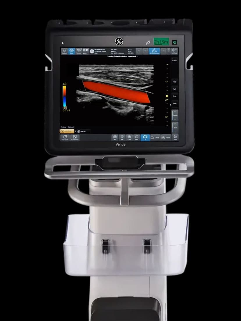
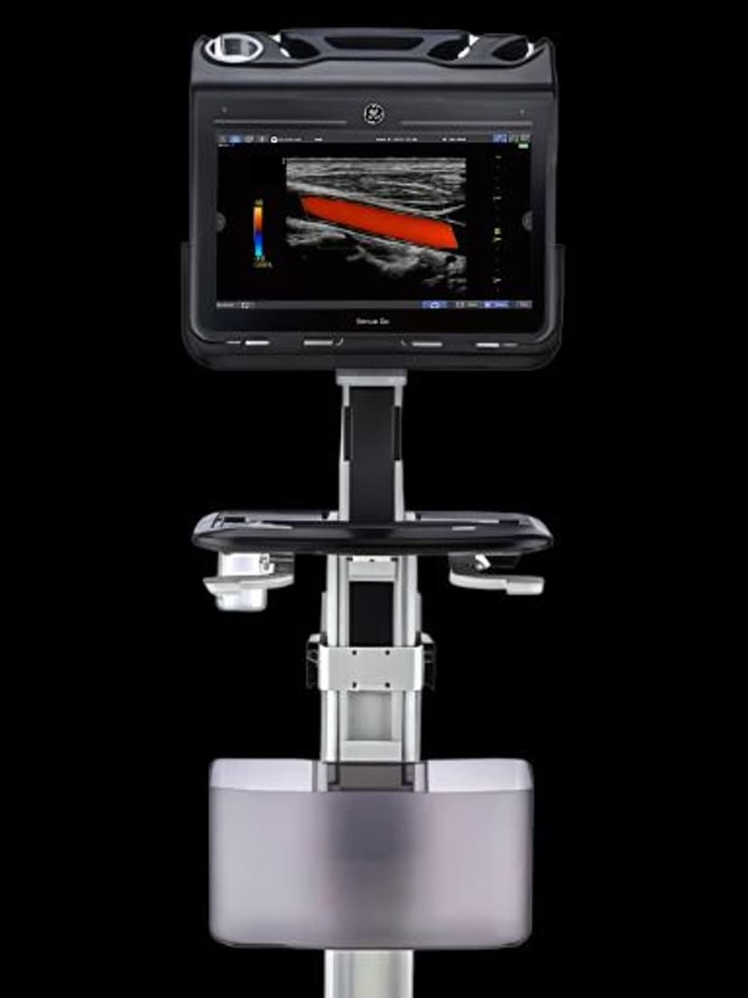
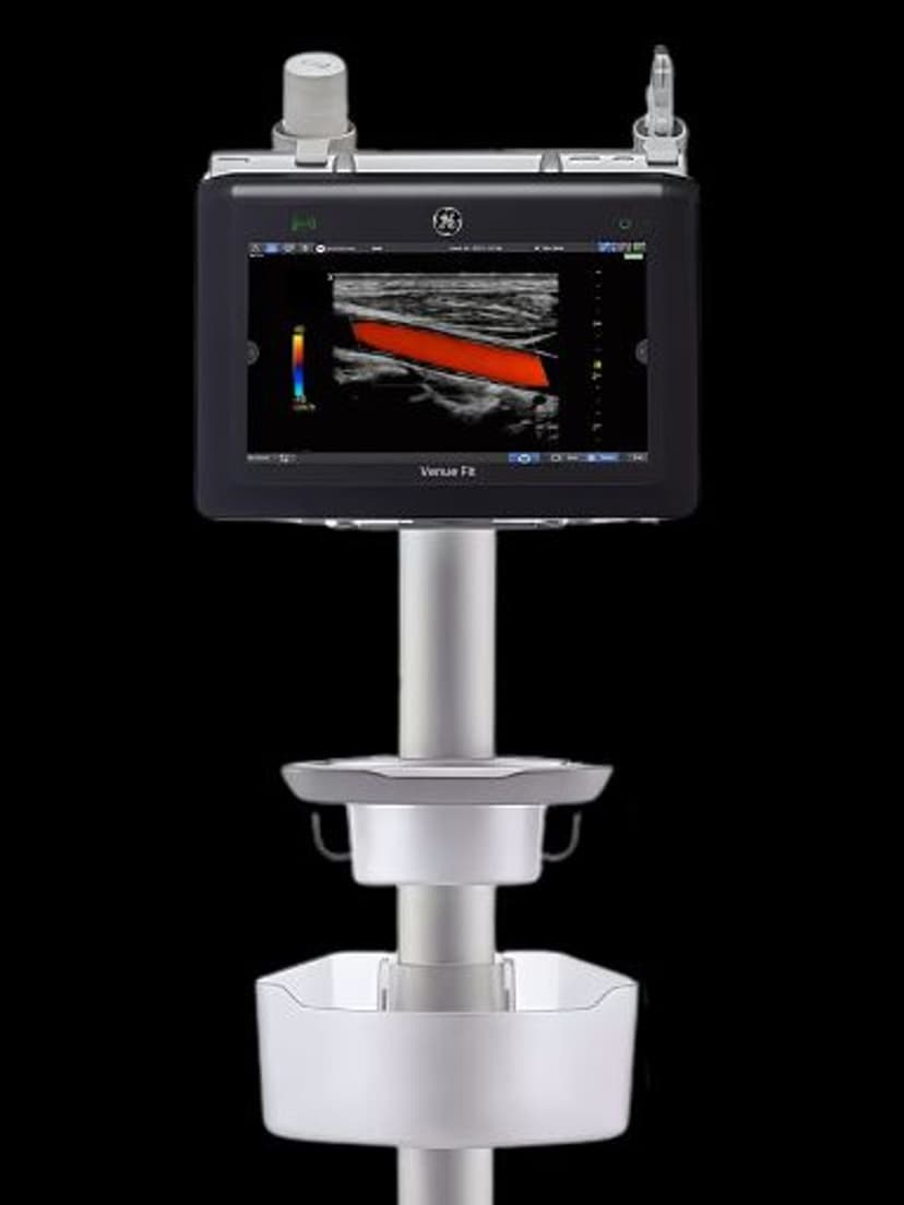
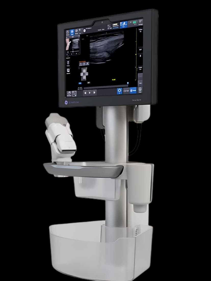
InterventionalAuto Vascular Access is an AI-powered feature on GE Venue systems that automatically identifies blood vessels during ultrasound-guided vascular access procedures. The system uses deep-learning algorithms to detect veins and arteries in the B-mode image, overlaying color-coded markers that distinguish vessels from surrounding tissue. This automated vessel identification reduces the time clinicians spend locating target vessels and improves the accuracy of needle placement, particularly for challenging patients with small, deep, or collapsed veins. Auto Vascular Access is designed for emergency departments, ICUs, and procedural suites where ultrasound-guided vascular access is performed by clinicians with varying levels of ultrasound experience.

Key Benefits
Why Auto Vascular Access matters
Instant vessel identification reduces search time
Auto Vascular Access highlights veins and arteries the moment the probe contacts the skin. Clinicians spend less time searching for the target vessel and more time executing the access procedure, improving first-attempt success rates and reducing patient discomfort.
Arterial vs. venous differentiation reduces cannulation errors
The AI distinguishes arteries from veins based on vessel characteristics, providing visual cues that help prevent accidental arterial cannulation during central venous access. This safety layer is particularly valuable for operators who perform the procedure infrequently.
Effective for difficult-access patients with small or deep veins
Obese patients, IV drug users, and patients with edema or hypovolemia often present with veins that are hard to locate by palpation or standard ultrasound. Auto Vascular Access detects and marks these vessels even when they are deep, small, or surrounded by edematous tissue.
Training support for clinicians building ultrasound-guided access skills
Emergency medicine residents, nursing staff, and other clinicians learning ultrasound-guided IV and central line placement benefit from the AI overlay as a real-time confirmation of their vessel identification. The visual feedback accelerates the learning curve for a procedure that traditionally requires significant hands-on experience.
About Auto Vascular Access
Ultrasound-guided vascular access has become standard practice for central venous catheter placement and increasingly common for peripheral IV insertion in difficult-access patients. The procedure requires the clinician to identify the target vessel, distinguish it from adjacent arteries, and guide the needle under real-time ultrasound visualization. For experienced sonographers, vessel identification is straightforward. But for emergency physicians, nurses, and other clinicians who perform vascular access intermittently, finding and confirming the target vessel can be the most time-consuming part of the procedure. Auto Vascular Access addresses this by applying AI vessel detection to the live B-mode image. The algorithm identifies circular or oval hypoechoic structures consistent with blood vessel cross-sections, evaluates their compressibility and pulsatility characteristics, and displays visual overlays that mark vessel locations and boundaries. The clinician sees the highlighted vessels immediately upon placing the probe, reducing the search time from seconds or minutes to instant confirmation. The feature also helps distinguish veins from arteries, which is critical for central line placement where inadvertent arterial cannulation is a serious complication. For training purposes, Auto Vascular Access gives less experienced operators a visual confirmation layer that builds confidence and correlates their developing anatomy recognition skills with AI-verified vessel identification.
Availability
Available on these systems
Our Partners


Want Auto Vascular Access in your practice?
Request a quote for a system that includes this feature.