
GE Ultrasound Feature
Bladder Volume Tool
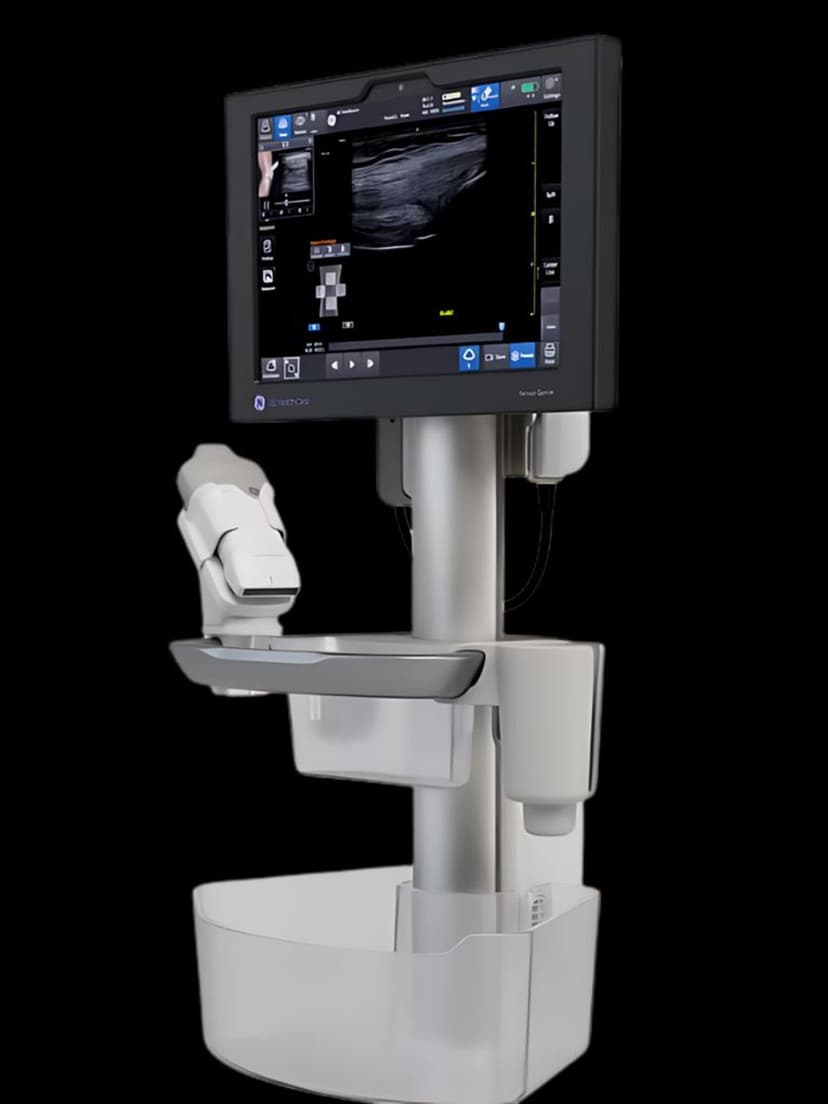
MeasurementThe Bladder Volume Tool guides clinicians through bladder volume measurement with on-screen reference images showing the correct transducer position and expected anatomy. The system uses edge-detection algorithms to automatically place calipers on the bladder walls and calculate volume in real time. Manual caliper adjustment is available for cases where bladder geometry is atypical. Designed for the GE Venue platform, this tool is built for point-of-care use in urology clinics, emergency departments, ICUs, and post-operative recovery areas where fast, reliable bladder assessment determines catheterization decisions.

Key Benefits
Why Bladder Volume Tool matters
On-screen guidance for non-specialist operators
Reference images and step-by-step visual prompts show clinicians where to place the transducer and what the target image should look like. Nurses, residents, and primary care physicians obtain accurate bladder volumes without dedicated ultrasound training.
Automated caliper placement reduces measurement variability
Edge-detection algorithms identify the bladder wall and position calipers at maximal dimensions. This removes the subjectivity of manual caliper placement, producing more consistent measurements across different operators and exam conditions.
Real-time volume display for immediate clinical decisions
The calculated volume appears on screen within seconds of image acquisition. In emergency and post-operative settings, this speed helps clinicians decide whether to catheterize, continue monitoring, or adjust fluid management without waiting for formal ultrasound reads.
Serial trending for urinary retention monitoring
Stored volume measurements can be compared across serial assessments to track bladder function recovery after surgery, monitor chronic urinary retention, or evaluate treatment effectiveness in patients with neurogenic bladder.
About Bladder Volume Tool
Bladder volume assessment is a high-frequency point-of-care exam performed by a wide range of clinicians, many of whom are not ultrasound specialists. The Bladder Volume Tool addresses this by combining guided scanning with automated measurement. On-screen reference images show the clinician where to place the transducer and what the expected image should look like in both transverse and sagittal orientations. This visual guidance helps non-specialist users acquire diagnostic-quality images without extensive ultrasound training. Once the image is acquired, edge-detection algorithms identify the bladder wall and automatically position calipers at the maximal dimensions. The system calculates volume using the ellipsoid formula and displays the result immediately. Clinicians can accept the automated measurement or manually adjust caliper positions when the bladder contour is irregular due to pelvic pathology, prior surgery, or incomplete distension. The tool integrates into the Venue system's clinical workflow protocols, allowing bladder volume measurement to be part of a standardized exam sequence. Results are stored and can be trended over serial assessments to monitor urinary retention patterns or post-operative recovery.
Availability
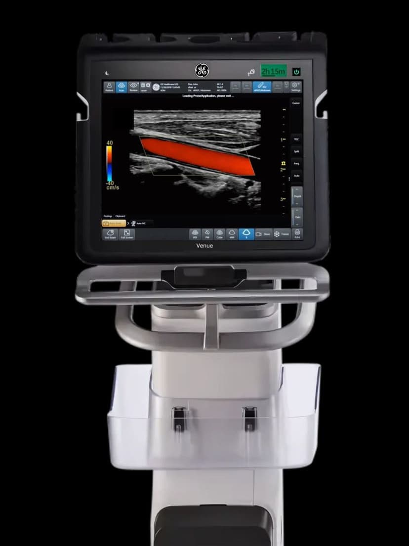
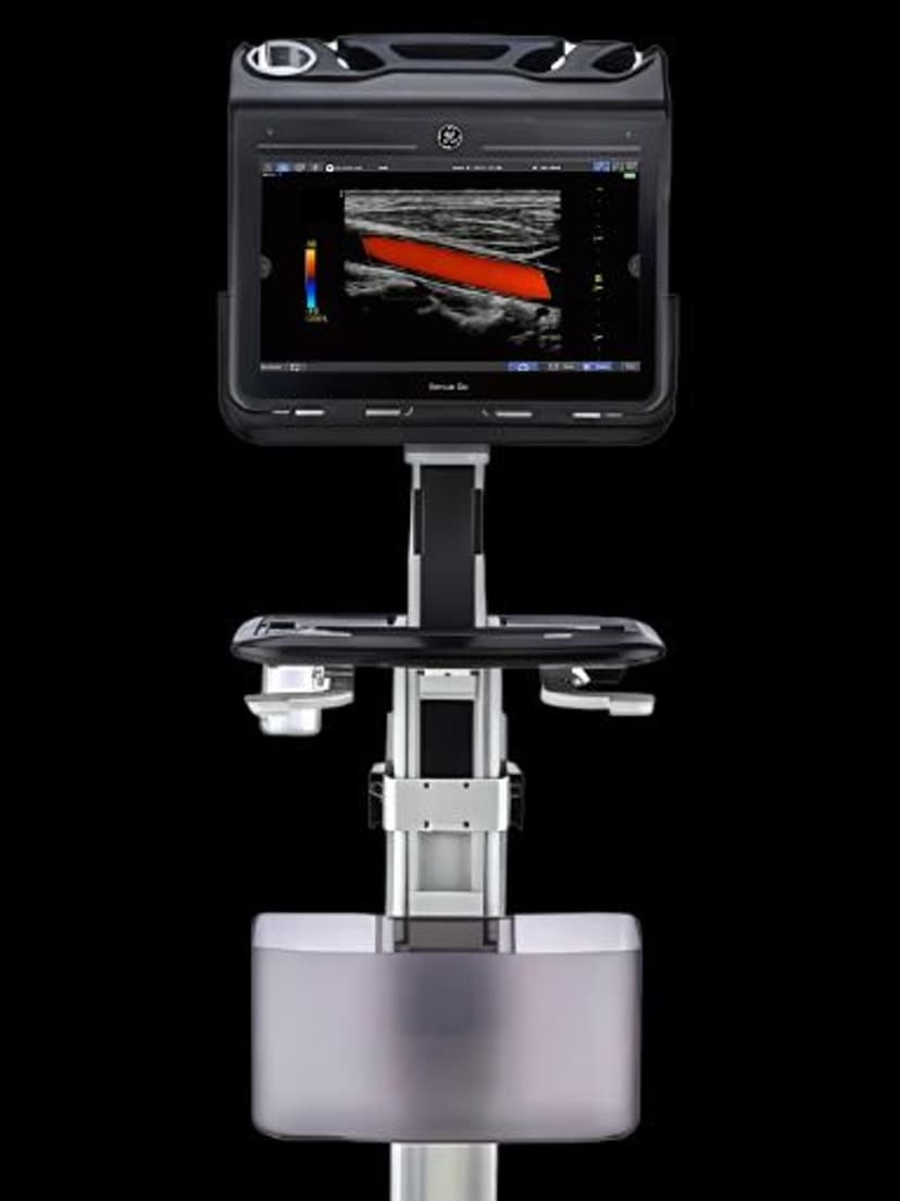
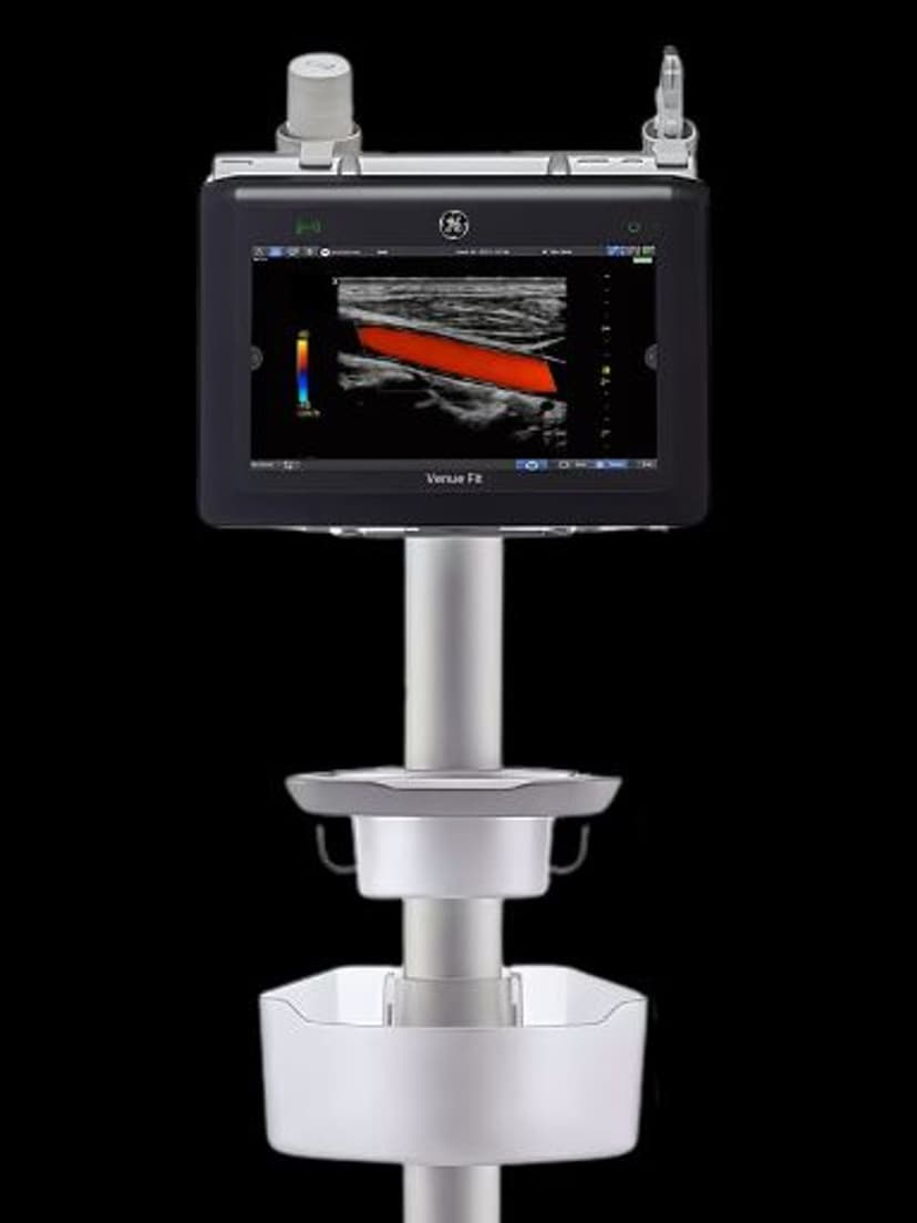
Available on these systems
Our Partners


Want Bladder Volume Tool in your practice?
Request a quote for a system that includes this feature.