
GE Ultrasound Feature
cNerve
AI & AutomationcNerve uses AI algorithms to automatically identify and highlight nerve structures on the ultrasound display during regional anesthesia procedures. The system detects nerve landmarks in real time and tracks them as the probe moves, reducing the time anesthesiologists spend searching for and confirming nerve locations before needle insertion. In clinical use, cNerve has identified nerve structures in up to 99% of cases. The feature works during both live scanning and stored clip review, so clinicians can verify nerve position after the procedure. Anesthesiologists performing peripheral nerve blocks benefit from faster nerve localization and increased confidence in needle placement relative to the target nerve.

Key Benefits
Why cNerve matters
Nerve detection in up to 99% of cases
The AI algorithm identifies nerve structures with high reliability across common block sites. This detection rate means fewer cases where the anesthesiologist must rely solely on indirect landmarks or loss-of-resistance techniques to locate the target nerve.
Continuous real-time tracking during needle advancement
cNerve tracks the nerve across frames as the probe and needle move, maintaining the visual overlay throughout the procedure. This persistent tracking reduces the risk of losing sight of the nerve during the most critical phase of the block.
Clear nerve visualization in difficult anatomy
Patients with high BMI, edema, or anatomical variants present challenges for manual nerve identification. The AI overlay highlights nerve boundaries that may be difficult to distinguish from surrounding fascial planes on standard B-mode imaging.
Post-procedure clip review for quality assurance
The detection algorithm applies to stored clips as well as live scanning. Anesthesia departments can review nerve block recordings to confirm proper needle-to-nerve positioning, supporting training programs and quality improvement initiatives.
About cNerve
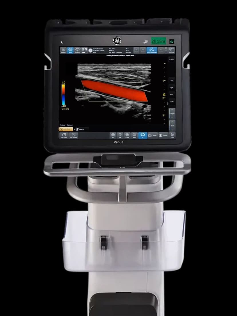
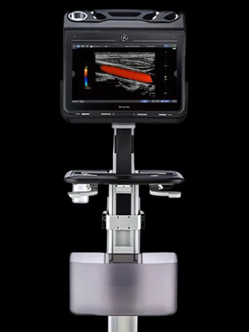
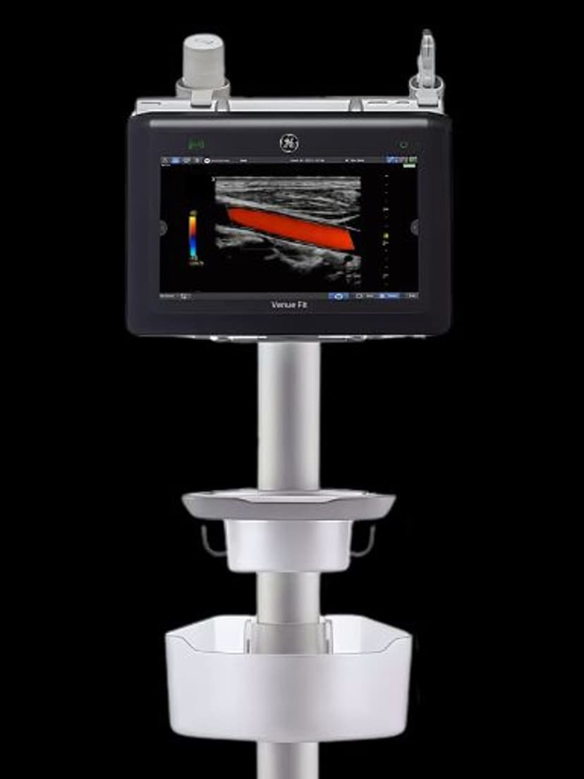
cNerve applies a trained AI model to B-mode ultrasound images to detect and segment nerve structures in real time. The algorithm identifies common peripheral nerve landmarks used in regional anesthesia, including the brachial plexus, femoral nerve, sciatic nerve, and interscalene targets. Once detected, the nerve boundary is highlighted with a color overlay on the live ultrasound image, and the system continuously tracks the structure as the operator adjusts probe position. This tracking persists across frames, reducing the need for the operator to repeatedly confirm anatomy during needle advancement. The feature also supports post-procedure review by applying the same detection algorithm to stored ultrasound clips. For anesthesiologists who perform high volumes of nerve blocks, cNerve reduces the cognitive load of manual nerve identification, particularly in patients with difficult anatomy such as those with high BMI or anatomical variants. The system is designed as a decision-support tool: it highlights structures for the clinician to confirm, not as a replacement for anatomical knowledge.
Availability
Available on these systems
Our Partners


Want cNerve in your practice?
Request a quote for a system that includes this feature.