
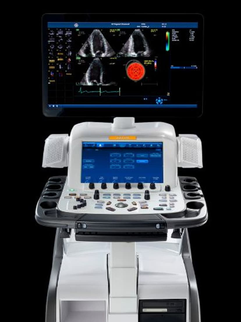
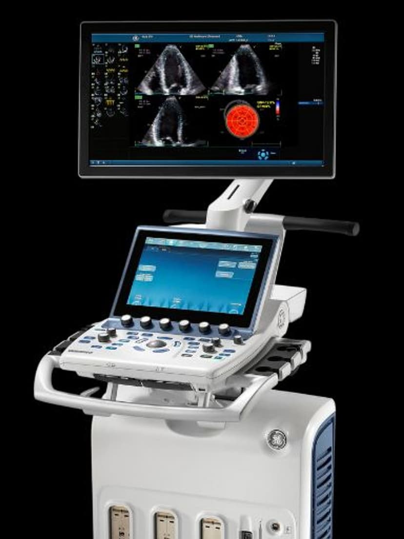
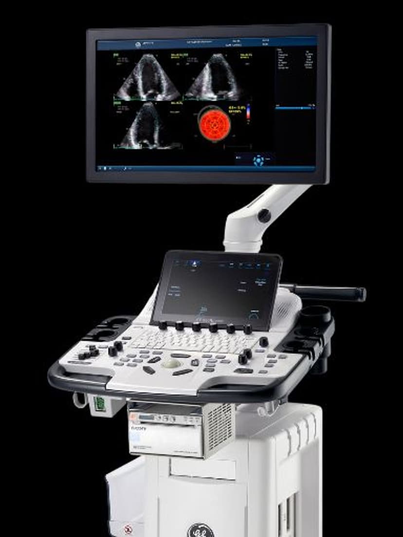
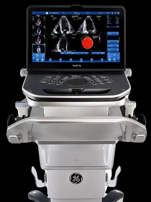
GE Ultrasound Feature
Color Flow
Imaging TechnologyColor Flow maps blood flow direction and relative velocity as a color overlay on the standard B-mode image. Red indicates flow toward the transducer, blue indicates flow away, and the color intensity reflects relative velocity. This real-time visualization allows clinicians to identify flow patterns, detect stenosis or occlusion, evaluate valvular regurgitation, and assess vascular patency without switching to a separate imaging mode. Color Flow is a fundamental Doppler tool available across GE ultrasound platforms, used in cardiac, vascular, abdominal, and OB/GYN applications wherever blood flow assessment is clinically relevant.

Key Benefits
Why Color Flow matters
Real-time flow direction and velocity mapping
Color Flow displays hemodynamic data directly on the anatomical image, so clinicians see flow patterns in context. This removes the guesswork of locating vessels or jets before switching to spectral Doppler for quantitative measurement.
Fast identification of stenosis, regurgitation, and occlusion
Abnormal flow patterns such as turbulent jets, aliasing at stenotic segments, or absent flow in thrombosed vessels appear immediately in the color overlay. This visual screening speeds the diagnostic process compared to relying on B-mode alone.
Adjustable sensitivity for low-flow to high-velocity applications
PRF, gain, and wall filter controls allow the operator to tune Color Flow sensitivity across a wide range of clinical scenarios, from low-velocity venous flow to high-velocity cardiac jets, using the same imaging tool.
Cross-specialty utility from cardiac to vascular to OB/GYN
Cardiologists use it for valve assessment, vascular specialists for carotid and peripheral evaluation, and OB/GYN providers for placental flow. A single feature serves multiple departments and exam types within the same practice.
About Color Flow
Color Flow Doppler operates by sampling multiple points along each scan line for frequency shifts caused by moving blood cells. The system assigns colors based on the direction and magnitude of the frequency shift relative to the transducer, then overlays this data on the grayscale B-mode image. The color box can be resized and repositioned to focus on the area of clinical interest, and parameters such as pulse repetition frequency, color gain, and wall filter are adjustable to optimize sensitivity for different flow conditions. Low-flow states require higher sensitivity settings, while high-velocity jets benefit from higher PRF to avoid aliasing. In cardiac imaging, Color Flow is essential for detecting and grading valvular regurgitation, visualizing intracardiac shunts, and assessing prosthetic valve function. In vascular imaging, it maps flow through carotid, peripheral, and abdominal vessels to identify stenosis, thrombosis, or aneurysmal flow patterns. OB/GYN applications include placental flow assessment and ovarian vascularity evaluation. Color Flow works with spectral Doppler modes (PW and CW) for quantitative velocity measurement after the color map identifies the area of interest.


Our Partners


Want Color Flow in your practice?
Request a quote for a system that includes this feature.