
GE Ultrasound Feature
Diagrams
WorkflowDiagrams provides a library of pre-loaded anatomical illustrations that can be displayed alongside or overlaid on the live ultrasound image during scanning. Operators select the relevant body region, and the system shows a labeled diagram of the expected anatomy for that area. This visual reference helps less experienced ultrasound users identify structures on screen by comparing the live image to the anatomical illustration. Diagrams also serves as a communication tool during bedside exams, allowing clinicians to show patients which structures are being examined and where findings are located relative to familiar anatomy.

Key Benefits
Why Diagrams matters
On-screen anatomical reference during scanning
Labeled illustrations display alongside the live ultrasound image, giving operators an immediate visual reference for identifying structures. This removes the need to consult separate anatomy references while scanning.
Faster structure identification for occasional users
Physicians who perform point-of-care ultrasound intermittently can match live anatomy to the diagram in real time. This reduces hesitation during bedside exams and supports confident structure identification without relying on memory alone.
Patient communication aid at the bedside
The anatomical diagrams give clinicians a visual tool for explaining findings to patients during the exam. Showing a labeled illustration alongside the ultrasound image makes findings more understandable for patients who cannot interpret ultrasound images on their own.
Built-in training resource for POCUS education
Residency programs and POCUS training courses can use Diagrams as an integrated teaching tool during hands-on scanning sessions. Trainees see the anatomical reference and the live image on the same screen, reinforcing anatomy recognition during real-time practice.
About Diagrams
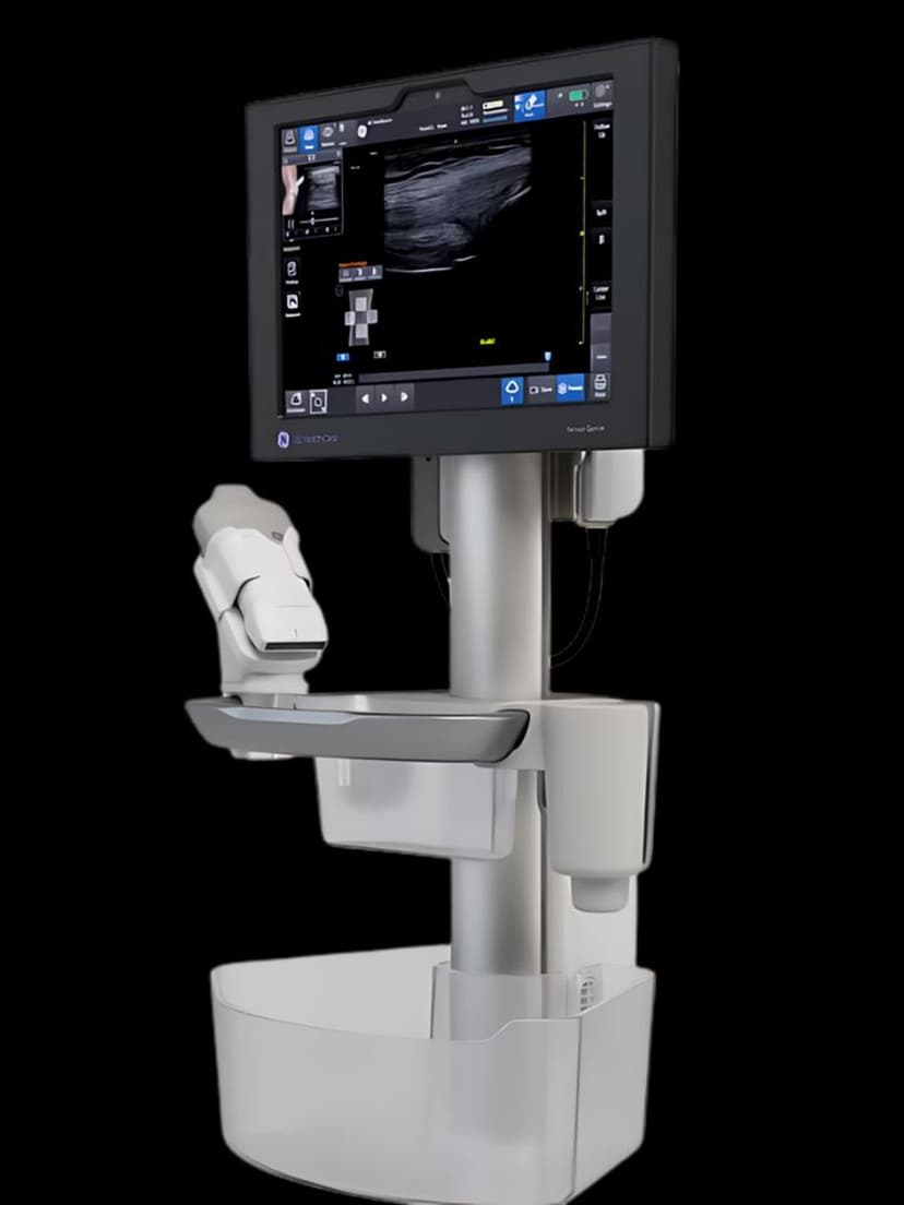
The Diagrams feature stores a library of anatomical illustrations organized by body region, including abdomen, thorax, vasculature, and musculoskeletal areas. When an operator selects a region, the corresponding diagram appears on the display, showing labeled organs, vessels, and landmarks relevant to that scanning area. The diagram can be viewed side by side with the live ultrasound image or overlaid for direct comparison. This reference system is designed for the GE Venue Sprint, a point-of-care system used in settings where operators may not perform ultrasound exams daily. In emergency departments, urgent care clinics, and primary care offices, the clinician performing the scan may be a physician whose primary specialty is not imaging. Diagrams bridges the gap between textbook anatomy and live ultrasound appearance, reducing the cognitive load of structure identification during bedside assessments. The feature also supports clinical education by providing an in-context anatomical reference during training sessions.
Availability
Available on these systems
Our Partners


Want Diagrams in your practice?
Request a quote for a system that includes this feature.