
GE Ultrasound Feature
Easy AFI 3.0
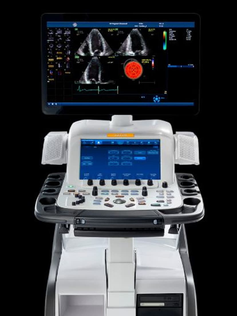
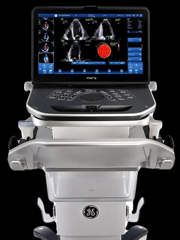
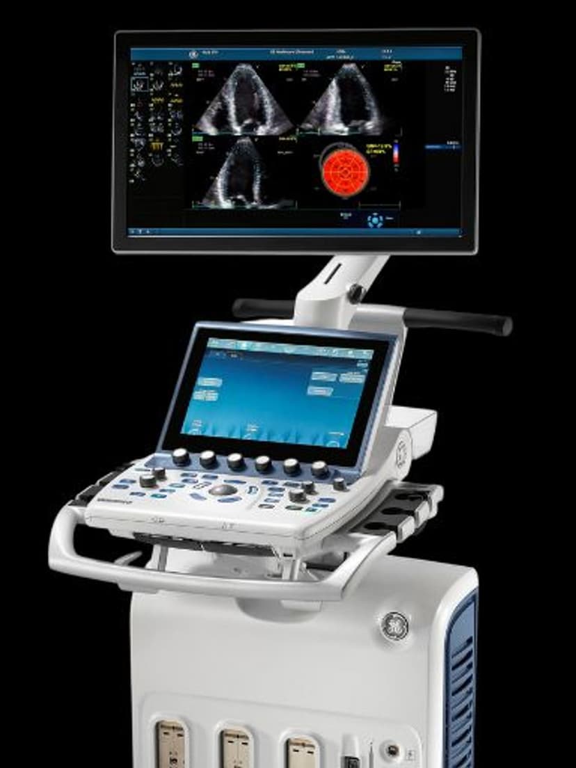
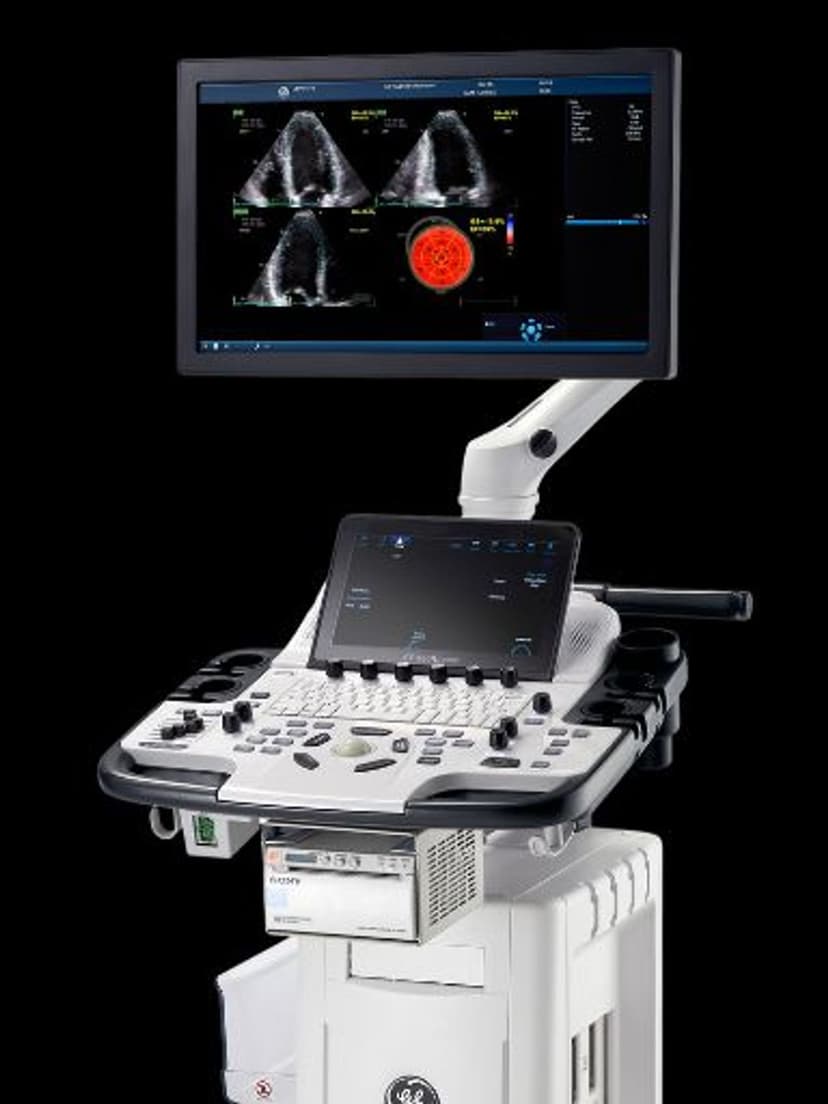
CardiacEasy AFI 3.0 automates myocardial strain analysis and ejection fraction measurement on GE Vivid-series echocardiography systems. The AI algorithm identifies apical views, detects endocardial borders, and tracks wall motion across the cardiac cycle to calculate global longitudinal strain (GLS) and ejection fraction without manual trace editing. Results appear in approximately 15 seconds. This removes the bottleneck of manual speckle tracking, which typically takes several minutes per view and varies between operators. Cardiologists, sonographers, and echo labs running high patient volumes benefit from faster, more reproducible strain data that supports clinical decisions in stress echo, chemotherapy cardiotoxicity monitoring, and routine cardiac function assessment.

Key Benefits
Why Easy AFI 3.0 matters
Strain and EF results in 15 seconds
The AI algorithm handles view recognition, border detection, and speckle tracking automatically. A process that manually takes 3–5 minutes per patient compresses to roughly 15 seconds, freeing time in echo labs running 15–20 studies per day.
Consistent results regardless of operator experience
By automating endocardial border detection and tracking, Easy AFI 3.0 reduces the inter-operator variability that makes manual strain measurements difficult to compare across follow-up exams or between different sonographers in the same lab.
Fewer manual steps than previous versions
Version 3.0 eliminates the manual view confirmation and border editing steps required in earlier AFI releases. Sonographers spend less time adjusting traces and more time acquiring quality images.
Reliable longitudinal tracking for cardiotoxicity monitoring
Oncology patients on cardiotoxic chemotherapy need serial strain measurements where small changes in GLS trigger dose adjustments. Easy AFI 3.0 delivers the reproducibility these protocols demand, so a 2-point GLS drop reflects actual cardiac change rather than measurement noise.
About Easy AFI 3.0
Easy AFI 3.0 builds on GE's Automated Function Imaging platform by adding AI-driven view recognition and border detection. The system acquires apical two-chamber, three-chamber, and four-chamber views, then automatically segments the left ventricle and applies speckle tracking to calculate segmental and global longitudinal strain. Ejection fraction is derived from the same dataset. The entire process, from image acquisition to bull's-eye strain map display, takes roughly 15 seconds. Previous versions required manual confirmation of endocardial borders and view selection. Version 3.0 reduces these manual steps, which directly lowers inter-operator variability. In busy echo labs, this means a sonographer with one year of experience can produce strain data comparable to an experienced operator. The feature supports stress echo protocols where time between stages is limited, and it provides reproducible longitudinal tracking for patients on cardiotoxic chemotherapy regimens where subtle changes in GLS trigger treatment modifications.


Our Partners


Want Easy AFI 3.0 in your practice?
Request a quote for a system that includes this feature.