
GE Ultrasound Feature
eFAST Diagram
WorkflowThe eFAST Diagram on GE Venue systems provides an on-screen visual map of all scanning zones in the Extended Focused Assessment with Sonography for Trauma protocol. During a trauma exam, the diagram displays the standard eFAST views: right upper quadrant, left upper quadrant, suprapubic, subxiphoid, and bilateral lung fields. Clinicians mark each zone as they complete it, ensuring full coverage of the protocol. This structured approach prevents missed views under the time pressure of emergency settings and keeps the exam organized for documentation.

Key Benefits
Why eFAST Diagram matters
Prevent missed views during high-pressure trauma exams
The on-screen diagram tracks which eFAST zones have been completed and which remain. In the controlled chaos of a trauma bay, this visual checklist prevents clinicians from accidentally skipping a critical view.
Label zones with a single keystroke
Clinicians mark each scanning zone as complete directly on the diagram without navigating menus or typing. This keeps the exam moving at the speed trauma cases demand.
Structured reporting for consistent documentation
Each completed zone links to its corresponding ultrasound images, creating an organized record of the exam. This structured approach supports quality review and meets documentation standards for trauma imaging.
Standardized eFAST protocol across skill levels
Residents and less experienced providers follow the same zone-by-zone workflow as senior clinicians. The diagram acts as a built-in protocol guide, reducing variability in exam completeness regardless of operator experience.
About eFAST Diagram
The eFAST protocol is a bedside ultrasound examination used in emergency departments and trauma bays to rapidly detect free fluid (blood) in the peritoneal and pericardial spaces and pneumothorax in the chest. The eFAST Diagram on GE Venue systems digitizes this protocol into an interactive on-screen guide. Each anatomical zone is represented on the body diagram, and clinicians can label completed views with a single keystroke, tracking their progress through the exam in real time. The diagram covers all standard eFAST views: Morrison's pouch (right upper quadrant), splenorenal recess (left upper quadrant), suprapubic/pelvic view, subxiphoid cardiac view, and bilateral anterior chest views for pneumothorax. By displaying which zones have been scanned and which remain, the diagram reduces the chance of skipping a view during a high-stress trauma evaluation. It also supports structured reporting by linking each completed view to the corresponding image, making post-exam documentation faster and more organized.
Availability
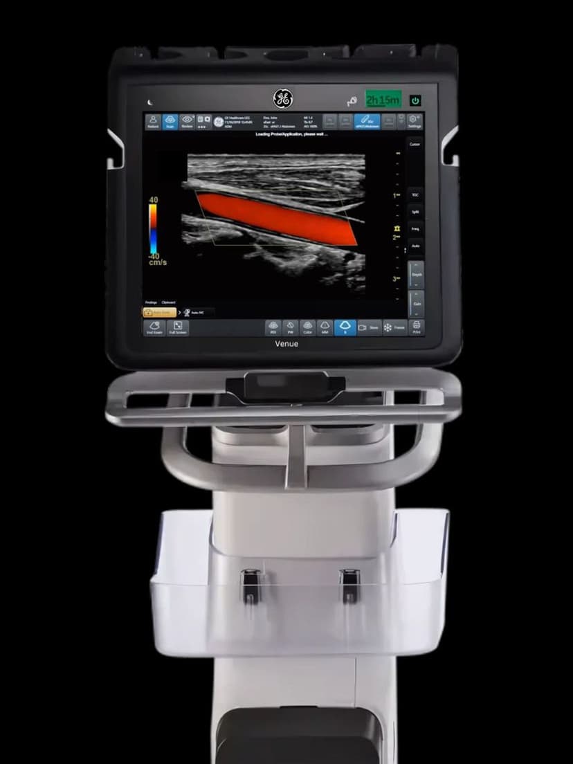
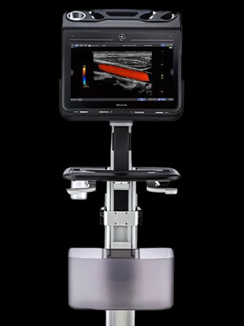
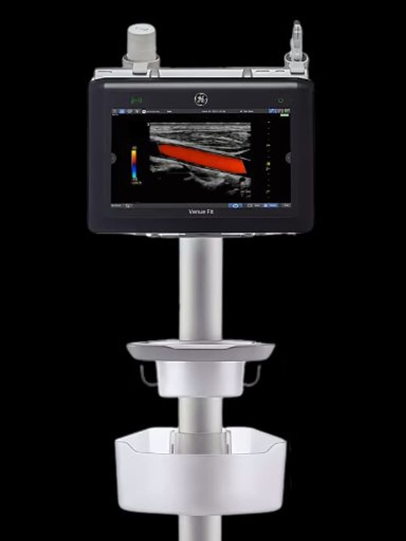
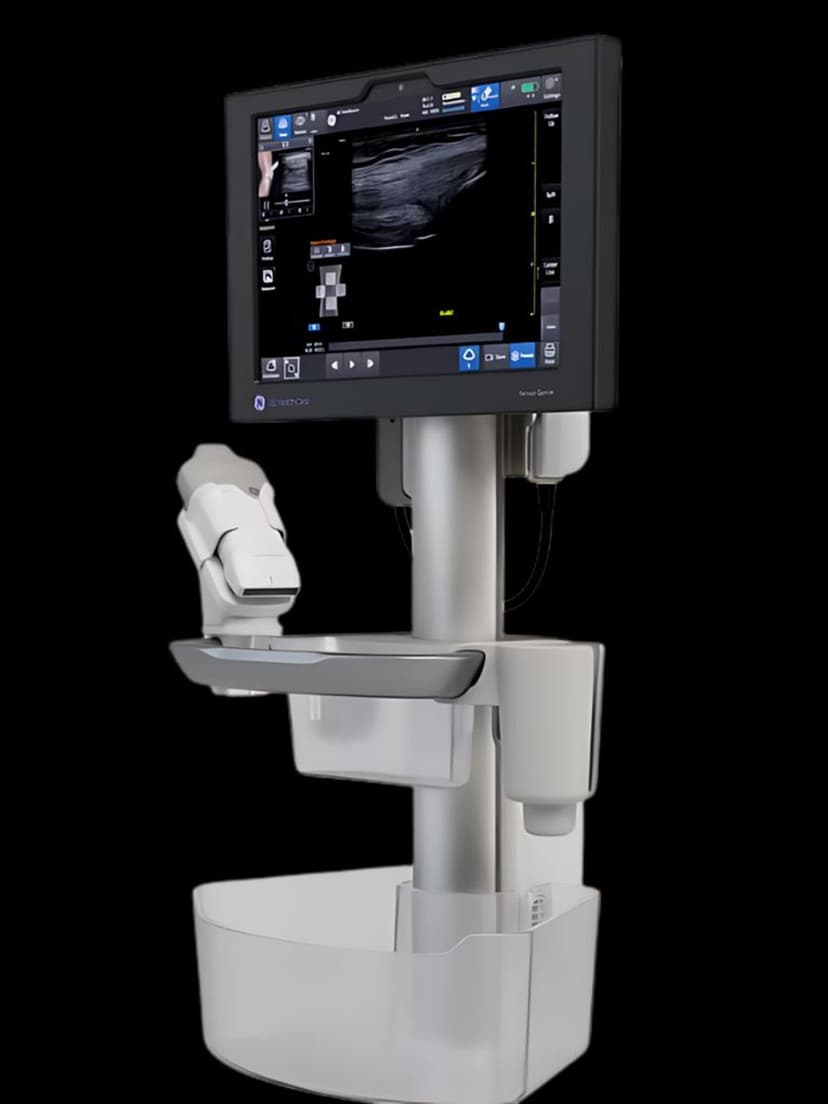
Available on these systems
Our Partners


Want eFAST Diagram in your practice?
Request a quote for a system that includes this feature.