
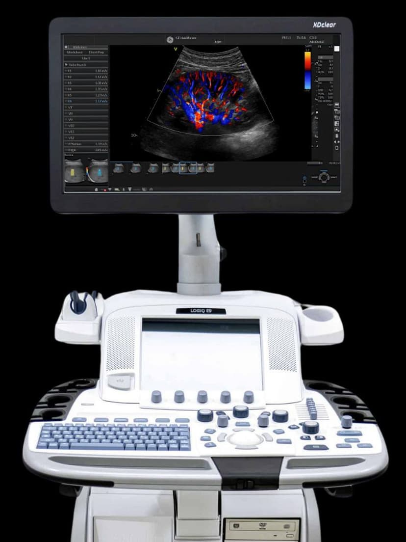
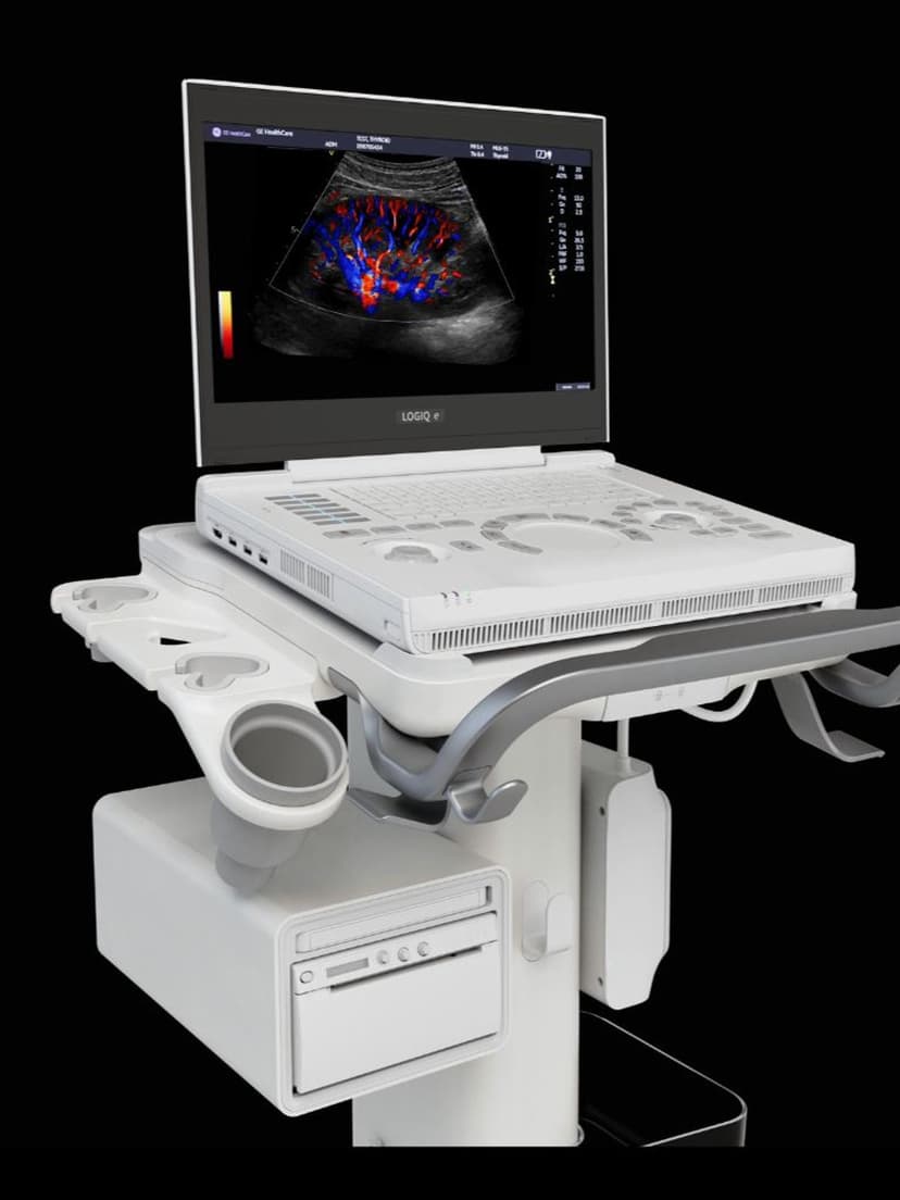
GE Ultrasound Feature
Elastography
MeasurementElastography measures tissue stiffness by sending shear waves through the target anatomy and tracking how the tissue deforms in response. Stiffer tissues (fibrotic liver, malignant tumors) propagate shear waves faster than softer tissues, and the ultrasound system converts these velocity differences into a quantitative stiffness map displayed in kilopascals (kPa) or meters per second (m/s). This gives clinicians an objective, non-invasive measure of tissue elasticity that can replace or reduce the need for biopsy in many clinical scenarios, particularly liver fibrosis staging where serial monitoring is common.

Key Benefits
Why Elastography matters
Non-invasive alternative to liver biopsy for fibrosis staging
Elastography provides a quantitative stiffness score that correlates with histological fibrosis stages (F0–F4). For chronic hepatitis and fatty liver disease monitoring, this eliminates the pain, risk, and cost of repeated percutaneous biopsies.
Improved breast lesion characterization
Adding tissue stiffness data to B-mode and Doppler findings improves the specificity of lesion assessment. Stiffer lesions are more likely malignant, and this additional information can help reduce unnecessary biopsies of BI-RADS 3 and 4a lesions.
Reproducible serial monitoring of disease progression
Quantitative stiffness values in kPa or m/s provide objective measurements that can be tracked over time. This is valuable for monitoring treatment response in hepatitis patients and tracking fibrosis progression in metabolic liver disease.
Multi-organ capability from a single tool
The same elastography technology applies across liver, breast, thyroid, MSK, and prostate applications. One feature extends the diagnostic reach of your LOGIQ system across multiple clinical specialties without additional hardware.
About Elastography
Shear wave elastography works by generating a mechanical push pulse using the ultrasound transducer's acoustic radiation force. This push creates shear waves that propagate laterally through the tissue. The ultrasound system tracks these waves at extremely high frame rates (thousands of frames per second) and calculates the shear wave velocity at each point in the region of interest. Because shear wave speed is directly related to tissue stiffness (Young's modulus), the system generates a color-coded elasticity map overlaid on the B-mode image. In hepatology, elastography provides a quantitative fibrosis score that correlates with histological staging (F0–F4), allowing clinicians to monitor disease progression or treatment response without repeated liver biopsies. For breast imaging, elastography adds a stiffness dimension to B-mode and Doppler findings, improving the specificity of lesion characterization and potentially reducing unnecessary biopsies of benign lesions. The technology also applies to thyroid nodule risk stratification, musculoskeletal tendon assessment, and prostate evaluation. GE's implementation on the LOGIQ platform includes 2D shear wave imaging with a confidence map that indicates measurement reliability, helping clinicians identify when results may be affected by patient factors like obesity, ascites, or breathing motion.


Our Partners


Want Elastography in your practice?
Request a quote for a system that includes this feature.