
GE Ultrasound Feature
FlexiView
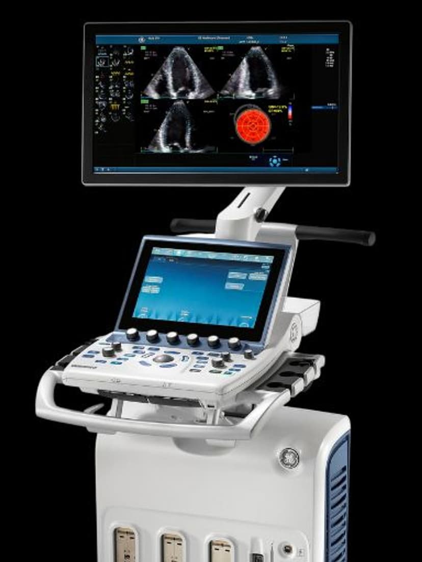
Imaging TechnologyFlexiView is a GE imaging tool that adjusts the angle and perspective of the ultrasound imaging plane in real time. Available on Vivid-series cardiac systems, FlexiView lets clinicians manipulate viewing angles during live scanning to visualize structures that are difficult to capture from a single fixed plane. This reduces the need to reposition the probe or rescan the patient when the initial view is suboptimal, particularly in echocardiography and abdominal imaging where acoustic windows can be limited.

Key Benefits
Why FlexiView matters
Capture diagnostic views from difficult acoustic windows
FlexiView adjusts the imaging plane angle in real time, so clinicians can visualize structures even when the acoustic window is narrow or obstructed. This is especially valuable in echocardiography where rib shadowing limits probe positioning.
Cut repeat scans by optimizing views in a single position
Rather than lifting the probe and repositioning, clinicians adjust the viewing angle on-screen. This reduces exam time and minimizes patient discomfort from repeated probe pressure.
Improved diagnostic confidence in complex anatomy
Multiple viewing perspectives from a single acquisition give clinicians more information to assess pathology. In cases where tissue differentiation is difficult, the additional angles help distinguish between structures that overlap in a standard plane.
More clinical value from 3D/4D volumes
In volumetric imaging modes, FlexiView lets operators navigate through reconstructed data to find planes that were not captured in the original sweep. This maximizes the diagnostic yield of each 3D/4D acquisition without additional scanning.
About FlexiView
FlexiView works by processing acquired image data and allowing the clinician to adjust the displayed viewing angle independently from the physical probe position. During a live exam, the operator can tilt, rotate, or shift the imaging plane to reveal anatomy that falls outside the standard scan plane. This is particularly useful in echocardiography, where intercostal spaces limit probe positioning, and in abdominal imaging, where bowel gas or patient body habitus can obstruct standard views. FlexiView reduces repeat scanning by giving clinicians the ability to optimize views from a single probe position. In 3D and 4D modes, FlexiView extends this flexibility further, allowing manipulation of reconstructed volumes to extract clinically relevant planes that would otherwise require additional acquisitions.
Availability
Available on these systems
Our Partners


Want FlexiView in your practice?
Request a quote for a system that includes this feature.