
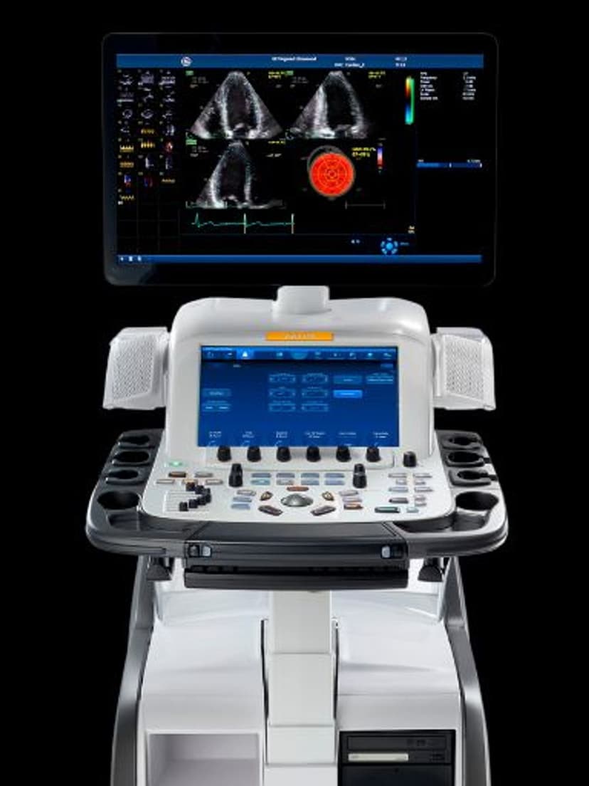
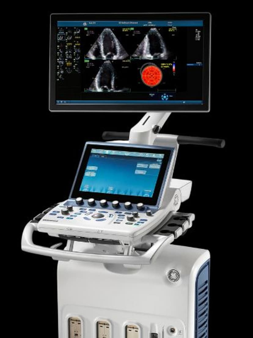
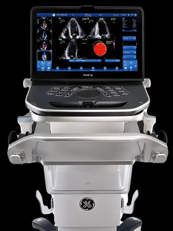
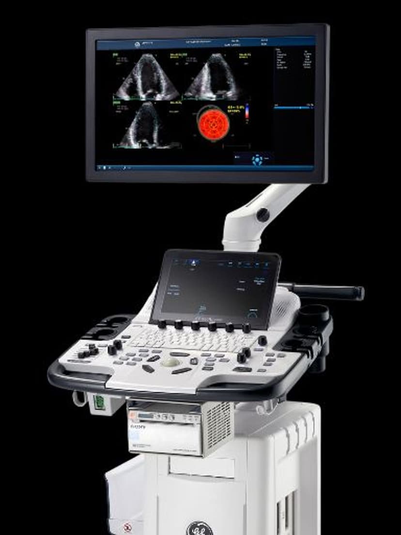
GE Ultrasound Feature
FlexiView Tool
Imaging TechnologyFlexiView Tool is a 3D/4D post-acquisition manipulation feature available on GE Vivid-series systems. After acquiring a volume dataset, clinicians can rotate, zoom, slice, and reposition the rendered image interactively to examine anatomy from any angle or plane. This eliminates the need to rescan when the original acquisition angle does not capture the ideal view. FlexiView is particularly useful in cardiac imaging for examining valve anatomy from non-standard planes, and in fetal imaging for clarifying structural anatomy when the initial scan position is suboptimal.

Key Benefits
Why FlexiView Tool matters
Diagnostic views without repeat scanning
FlexiView lets clinicians reposition and re-slice stored volume data to find the optimal viewing angle. If the original acquisition missed an ideal plane, the clinician retrieves it from the volume dataset rather than calling the patient back.
Fewer repeat scans and improved patient throughput
When a suboptimal scan angle would previously require repositioning the probe and rescanning, FlexiView provides the needed view from existing data. This cuts time per exam and reduces patient discomfort from extended scanning sessions.
Better visualization of complex cardiac and fetal anatomy
Rotating and slicing volume data reveals structures that are difficult to image in a single 2D plane. Valve morphology, septal anatomy, and fetal limb positioning become clearer when examined from multiple angles within the same dataset.
Separation of acquisition from interpretation
A sonographer acquires the 3D/4D volume, and a physician manipulates it later using FlexiView. This workflow model supports training programs where less experienced staff perform acquisitions and senior clinicians extract diagnostic information from the stored volumes.
About FlexiView Tool
FlexiView Tool operates on stored 3D and 4D volume datasets, giving clinicians full control over the viewing angle, zoom level, and slice plane after the scan is complete. The tool renders the volume data in real time, so adjustments appear immediately on screen without processing delay. In cardiac applications, FlexiView lets echocardiographers examine valve leaflets, septal defects, and chamber geometry from angles that may be difficult to achieve with probe positioning alone. In obstetric and gynecological imaging, the tool supports detailed fetal anatomical assessment by allowing the sonographer to rotate the volume to visualize structures obscured by fetal position. FlexiView reduces operator dependency because a less experienced sonographer can acquire the volume dataset, and a more experienced clinician can later manipulate the data to extract the diagnostic views needed. This separation of acquisition from interpretation is a practical advantage in training environments and in practices where scanning and reading are performed by different staff members.
Availability
Available on these systems
Our Partners


Want FlexiView Tool in your practice?
Request a quote for a system that includes this feature.