
GE Ultrasound Feature
HDlive
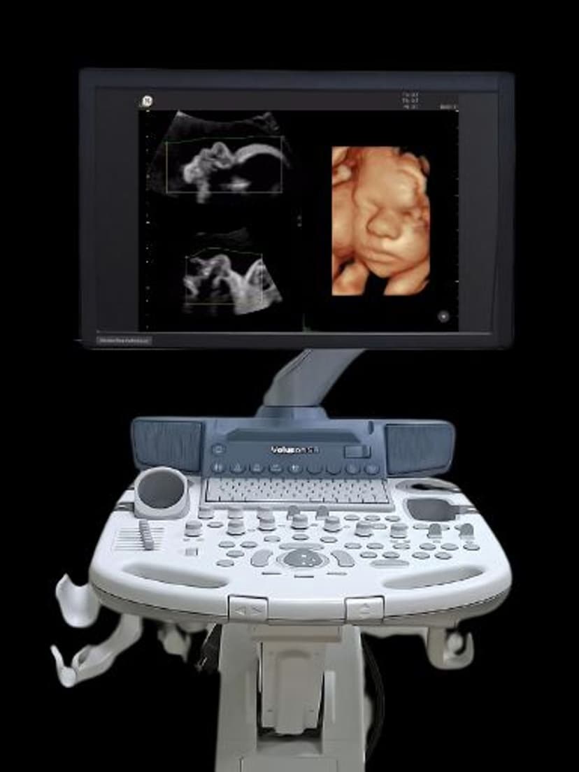
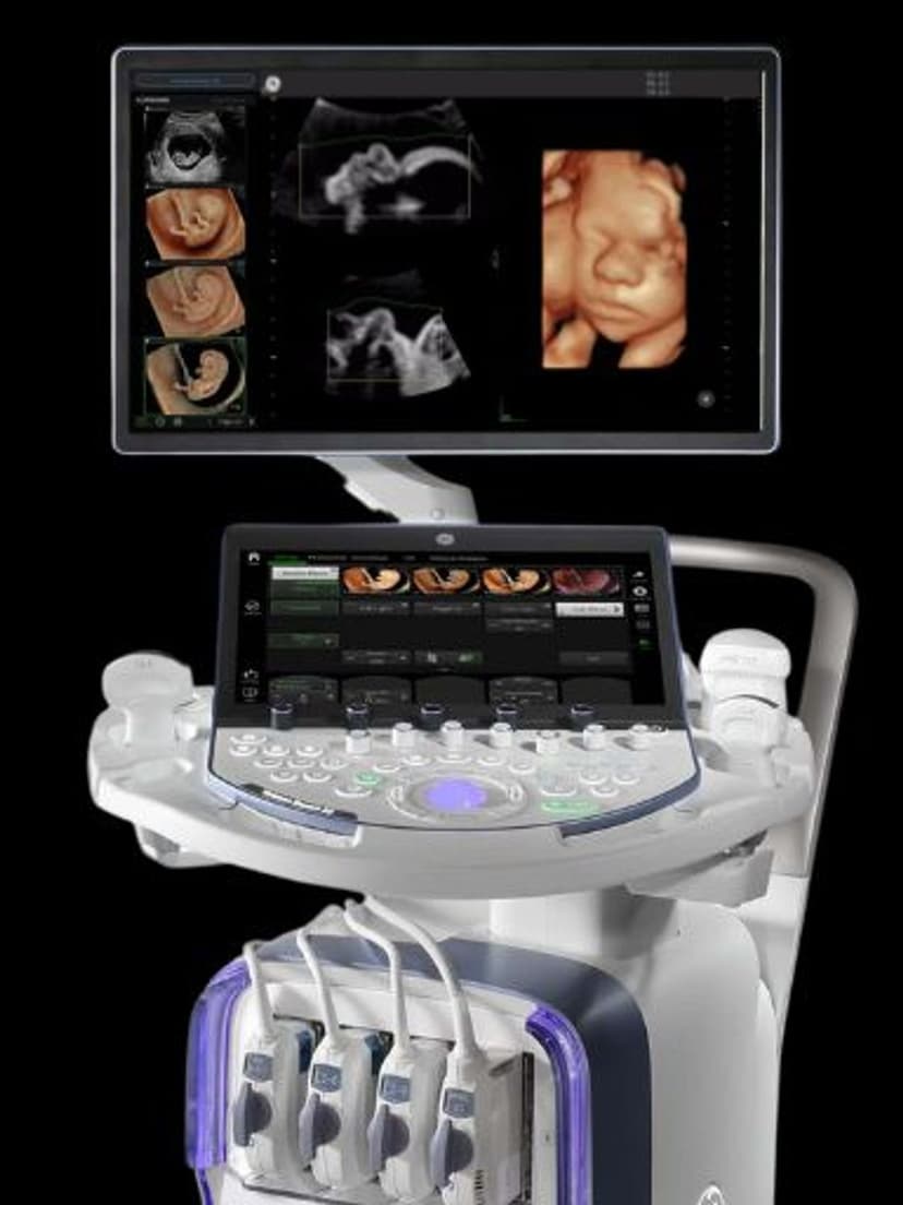
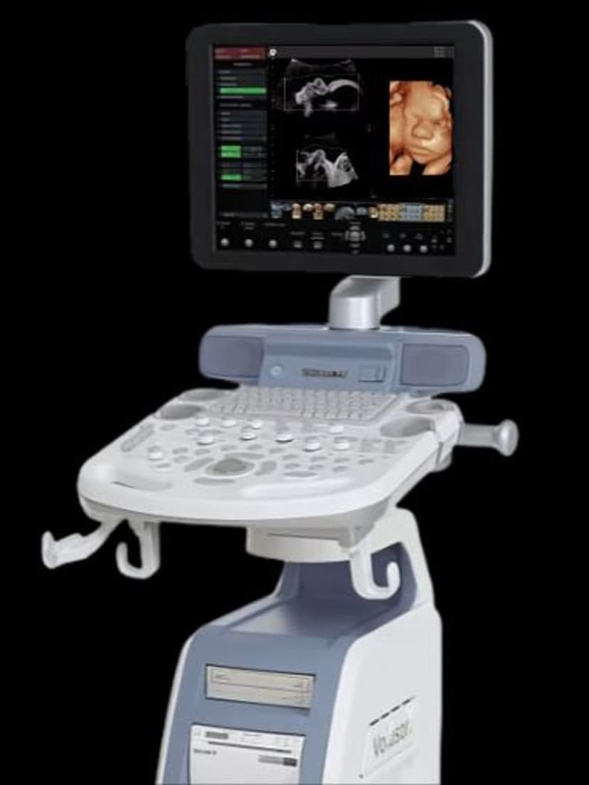
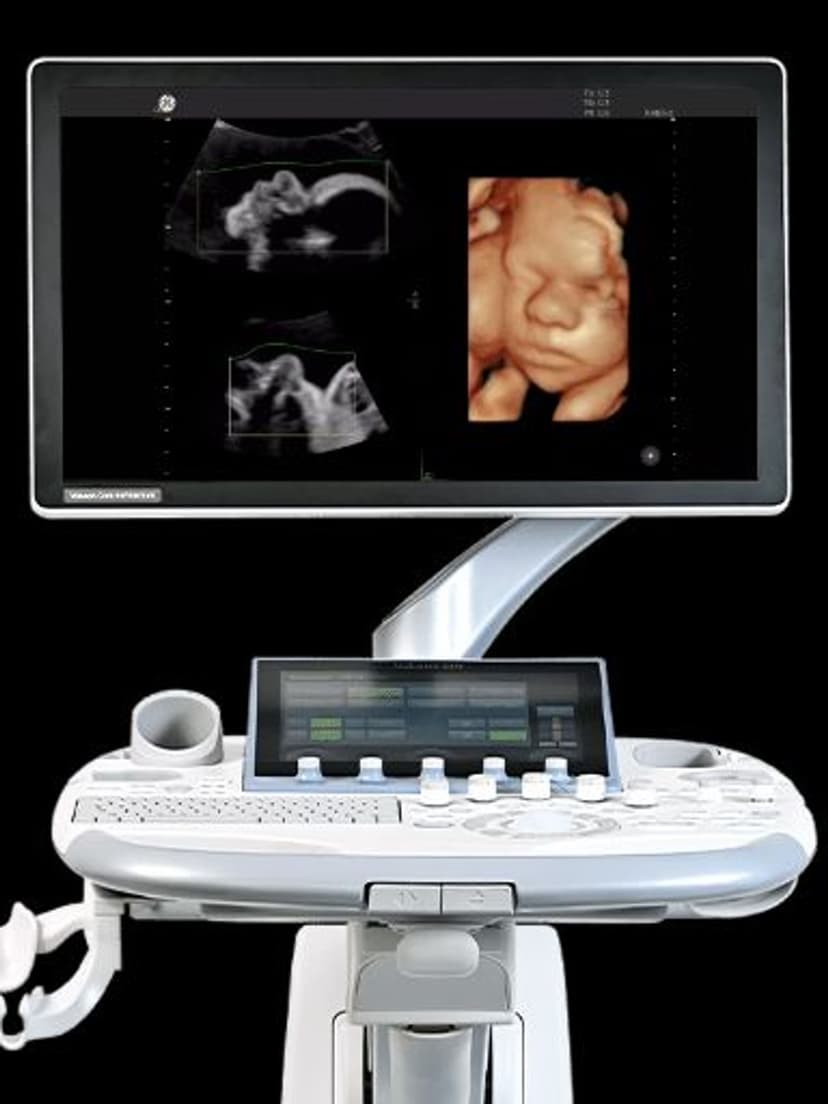
Imaging TechnologyHDlive applies a virtual, adjustable light source to 3D/4D ultrasound volume data, producing images with natural skin tones, realistic shadows, and depth perception that surpasses conventional surface rendering. The user positions the light at different angles to reveal facial features, surface anatomy, and structural details with clarity comparable to a photograph. HDlive is the standard for fetal face visualization in OB/GYN practices and also improves assessment of uterine anomalies, endometrial pathology, and adnexal masses in gynecological imaging.

Key Benefits
Why HDlive matters
Photorealistic fetal imaging with adjustable lighting
Position the virtual light source at any angle to reveal facial features, finger details, and surface anatomy. The resulting images show natural skin tones and shadows that parents immediately recognize as their baby.
Improved detection of surface anomalies
Natural shadowing from the virtual light source reveals facial clefts, limb abnormalities, and surface defects that flat 3D rendering can miss. Clinicians gain additional diagnostic confidence when assessing fetal anatomy.
Practice differentiation that drives referrals
HDlive images are visually distinct from standard 3D ultrasound. OB/GYN practices offering HDlive report higher patient satisfaction scores, increased word-of-mouth referrals, and stronger social media presence from shared images.
Gynecological visualization beyond obstetrics
HDlive renders uterine anomalies, endometrial polyps, fibroids, and adnexal masses with intuitive 3D depth that aids clinical interpretation. The same rendering technology that produces fetal images also improves assessment of gynecological pathology.
About HDlive
Traditional 3D ultrasound rendering produces flat, monochromatic surface images that can be difficult for patients and sometimes clinicians to interpret. HDlive changes this by simulating how light interacts with tissue surfaces. The rendering engine calculates light attenuation and scattering based on tissue density, creating natural shadows that reveal depth and contour. The virtual light source can be repositioned in real time, allowing the operator to highlight different aspects of the anatomy from multiple illumination angles. Speckle reduction algorithms and advanced beamforming work together to minimize noise artifacts, producing high-resolution volume images. HDlive supports imaging throughout all trimesters. In early pregnancy, it visualizes embryonic structures and early anatomical development. In second and third trimester, it provides detailed fetal face, limb, and surface anatomy assessment. Clinically, the enhanced depth perception improves detection of facial clefts, limb anomalies, and neural tube defects. For practice growth, HDlive images drive patient satisfaction, social media sharing, and referrals from expectant parents who value the bonding experience.

Our Partners


Want HDlive in your practice?
Request a quote for a system that includes this feature.