
GE Ultrasound Feature
High Res PDI
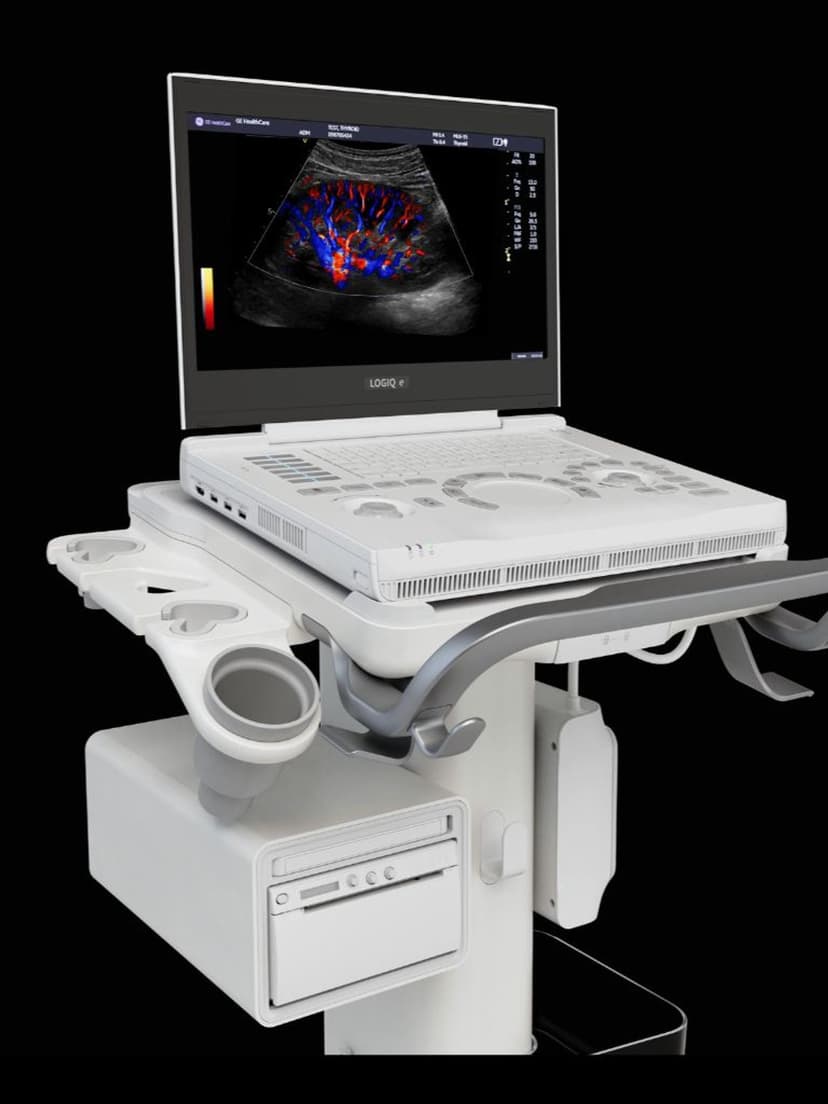
Imaging TechnologyHigh Res PDI is GE's high-resolution Power Doppler Imaging mode, available on LOGIQ systems. Unlike conventional color Doppler, which displays blood flow direction and velocity, Power Doppler detects the amplitude of Doppler signals, making it more sensitive to slow flow in small vessels. High Res PDI adds spatial resolution to this sensitivity, producing detailed maps of microvascular perfusion in organs like the kidney, liver, and thyroid. Clinicians use it to evaluate tissue vascularity, detect subtle flow in tumor neovasculature, and assess perfusion in transplanted organs where small-vessel flow is clinically significant.

Key Benefits
Why High Res PDI matters
Detects blood flow in vessels too small for standard color Doppler
High Res PDI picks up slow, low-volume flow in capillary-level vasculature that falls below the detection threshold of conventional color Doppler. This matters for evaluating organ perfusion and identifying hypervascularity in suspicious masses.
Angle-independent flow mapping for more complete vascular assessment
Because Power Doppler measures signal amplitude rather than frequency shift, it does not suffer from angle dependency. Clinicians get consistent flow detection regardless of the Doppler angle, reducing the need to reposition the probe to optimize the color signal.
Sharper spatial resolution distinguishes adjacent vascular structures
The high-resolution processing separates individual small vessels that would blur together in standard Power Doppler mode. This spatial precision is useful for mapping tumor vascularity and differentiating vascular from avascular structures within complex masses.
Cross-specialty utility from vascular to OB/GYN to MSK
High Res PDI applies across multiple clinical domains. Vascular specialists use it for peripheral perfusion, radiologists for organ assessment, OB/GYNs for placental and adnexal evaluation, and rheumatologists for synovial inflammation. One imaging mode serves multiple referral patterns.
About High Res PDI
Standard color Doppler imaging encodes blood flow velocity and direction using frequency shift data, but it has a minimum detectable velocity threshold that misses slow flow in small vessels. Power Doppler Imaging works differently: it measures the total integrated power of the Doppler signal rather than the frequency shift, making it angle-independent and more sensitive to low-velocity flow. High Res PDI on GE LOGIQ systems adds higher spatial resolution to Power Doppler by using optimized pulse sequences and signal processing that preserve fine vascular detail. The result is a color overlay that maps blood flow presence and relative intensity across the scan area, revealing microvascular structures that standard color Doppler cannot resolve. Clinical applications include evaluating renal perfusion, detecting hypervascularity in thyroid nodules, assessing synovial inflammation in joints, and monitoring transplant organ perfusion. In OB/GYN, High Res PDI helps visualize placental blood flow and assess adnexal mass vascularity. The mode is particularly valuable when the clinical question is about whether blood flow is present in a structure, rather than its velocity or direction.
Availability
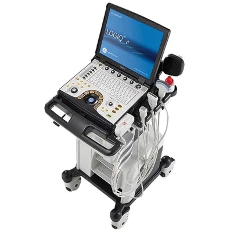
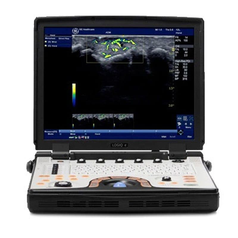
Available on these systems
Our Partners


Want High Res PDI in your practice?
Request a quote for a system that includes this feature.