
GE Ultrasound Feature
Lesion Characterization
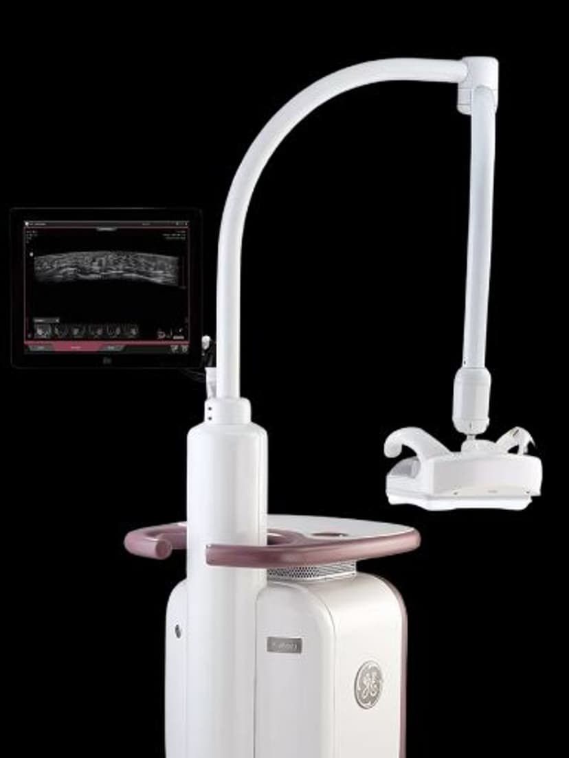
Imaging TechnologyLesion Characterization on the GE Invenia ABUS (Automated Breast Ultrasound System) provides structured analysis of breast lesion features to support BI-RADS classification. The system evaluates lesion shape, margin regularity, echogenicity patterns, posterior acoustic features, and internal vascularity, then presents these findings in a standardized format. Radiologists reading ABUS volumes use Lesion Characterization to document their assessment consistently and support diagnostic confidence when differentiating benign masses from suspicious findings requiring biopsy.

Key Benefits
Why Lesion Characterization matters
Structured BI-RADS-aligned lesion analysis
Lesion Characterization walks through each ACR BI-RADS descriptor systematically: shape, margin, echogenicity, posterior features, and vascularity. This structured approach ensures no morphological feature is overlooked during the read, reducing the risk of incomplete assessment.
Standardized reporting across radiologists and sites
When multiple radiologists read ABUS volumes across different locations, interpretation variability can affect callback rates and biopsy recommendations. Lesion Characterization enforces a consistent evaluation framework, so the same finding receives the same structured assessment regardless of who reads it.
Morphological detail supports biopsy decision-making
By presenting a complete set of lesion features in one view, Lesion Characterization gives radiologists the information they need to assign BI-RADS categories with confidence. Clearer characterization of benign-appearing features (oval shape, circumscribed margins, posterior enhancement) can reduce unnecessary biopsy recommendations.
Complements ABUS screening for dense breast tissue
ABUS excels at detecting lesions in dense breast tissue where mammography sensitivity drops. Lesion Characterization adds the morphological assessment layer needed to determine which ABUS-detected findings warrant further workup, completing the screening-to-diagnosis workflow without requiring a separate handheld ultrasound exam.
About Lesion Characterization
Breast lesion characterization follows the ACR BI-RADS lexicon, which defines specific morphological features that correlate with malignancy risk. Lesion Characterization on the Invenia ABUS systematizes this assessment by guiding the radiologist through each BI-RADS descriptor category. For shape, the system helps distinguish between oval (typically benign), round, and irregular (higher suspicion) morphologies. Margin analysis identifies circumscribed, indistinct, angular, microlobulated, or spiculated borders. Echogenicity classification ranges from anechoic (simple cyst, almost always benign) through hyperechoic, with complex patterns receiving additional scrutiny. The system also evaluates posterior acoustic features (enhancement, shadowing, or combined patterns) and, when Color Doppler data is available, internal vascularity. All findings populate a structured report template that aligns with BI-RADS categories 2 through 5, supporting the radiologist's final assessment and recommendation. For ABUS screening programs, standardized lesion characterization is especially important because the radiologist may be reading volumes from multiple technologists across different locations. Consistent characterization criteria reduce variability in how lesions are described and categorized, leading to more uniform biopsy recommendations and callback rates.
Availability
Available on these systems
Our Partners


Want Lesion Characterization in your practice?
Request a quote for a system that includes this feature.