
GE Ultrasound Feature
Lesion Detection
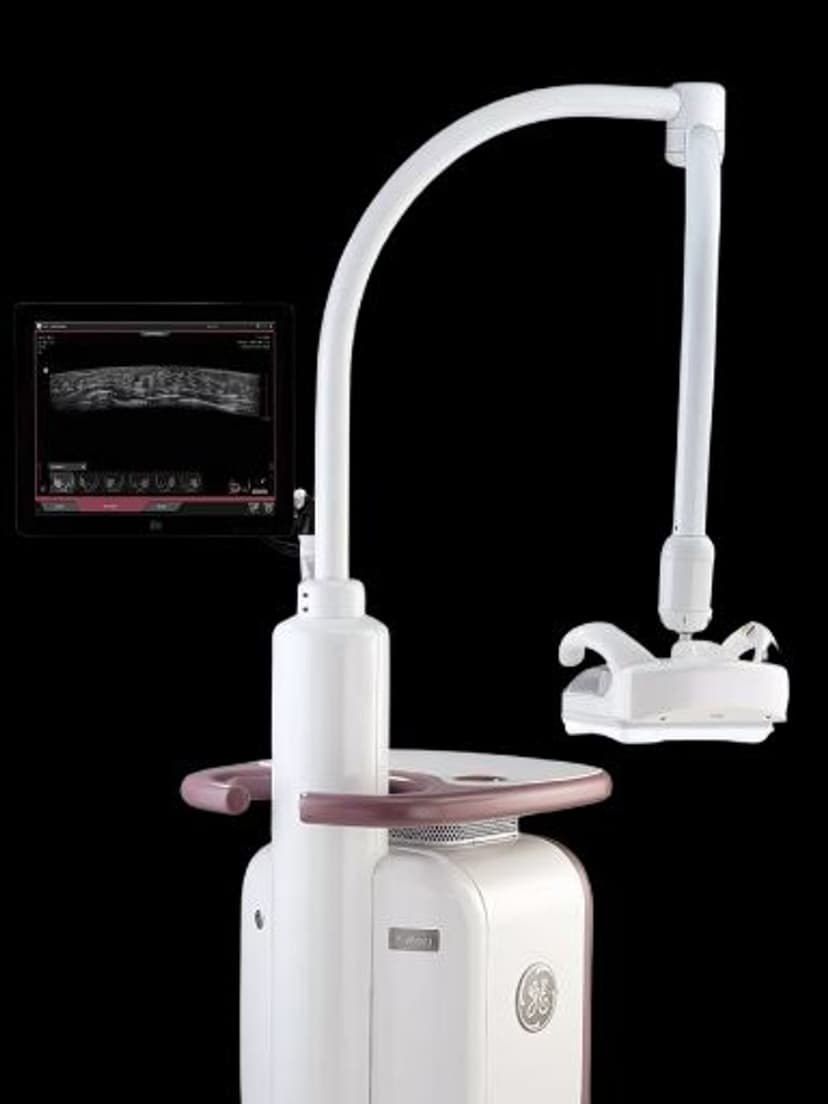
AI & AutomationLesion Detection applies deep-learning algorithms to automatically identify and trace lesion boundaries during ultrasound exams. When a clinician identifies a suspected lesion, the system segments its outline and calculates dimensions, removing the manual tracing and measurement steps. The technology works across multiple organ systems including breast, liver, and thyroid imaging, and integrates with contrast-enhanced ultrasound (CEUS) for improved visualization of vascularized lesions. On the GE Invenia ABUS platform, Lesion Detection supports automated breast screening by flagging suspicious findings for radiologist review.

Key Benefits
Why Lesion Detection matters
Consistent lesion measurements for reliable follow-up
Deep-learning segmentation produces the same lesion boundaries regardless of which operator performs the scan. Serial measurements reflect actual lesion changes rather than inter-operator tracing differences, giving oncologists more reliable data for treatment decisions.
Automated tracing replaces manual caliper work
The system identifies lesion margins and calculates dimensions without manual boundary tracing. In exams with multiple lesions, this automation saves several minutes per study and reduces the repetitive strain of fine caliper placement.
Multi-organ support across breast, liver, and thyroid
A single detection platform works across the organ systems where lesion measurement is most common. Practices do not need separate specialty tools for each application, and the measurement approach stays consistent across clinical contexts.
CEUS integration for vascularized lesion characterization
When used with contrast-enhanced ultrasound, Lesion Detection tracks enhancement patterns through wash-in and wash-out phases. This time-based vascular data adds a characterization layer beyond simple size measurement, supporting more confident diagnostic assessment of liver lesions.
About Lesion Detection
Manual lesion measurement in ultrasound requires the operator to trace the lesion boundary by hand and place calipers for diameter measurements. This process is subjective and varies between operators, which creates inconsistency in serial measurements used to track lesion growth over time. Lesion Detection replaces manual tracing with deep-learning segmentation that identifies lesion margins at the pixel level. The algorithm produces consistent boundaries regardless of who performs the exam, which makes follow-up comparisons more reliable. In breast imaging on the Invenia ABUS, the system processes automated whole-breast ultrasound volumes and highlights regions of interest for the radiologist, serving as a computer-aided detection layer that reduces the risk of overlooked findings in dense breast tissue. For liver and thyroid applications, Lesion Detection works in both standard B-mode and contrast-enhanced modes. During CEUS exams, the system can track lesion enhancement patterns over time, providing wash-in and wash-out data that supports lesion characterization. Clinicians can adjust the auto-generated boundaries when the algorithm's initial segmentation needs refinement, maintaining physician control over the final measurement. The consistent measurement approach is particularly valuable for oncology follow-up, where small changes in lesion dimensions guide treatment decisions.
Availability
Available on these systems
Our Partners


Want Lesion Detection in your practice?
Request a quote for a system that includes this feature.