
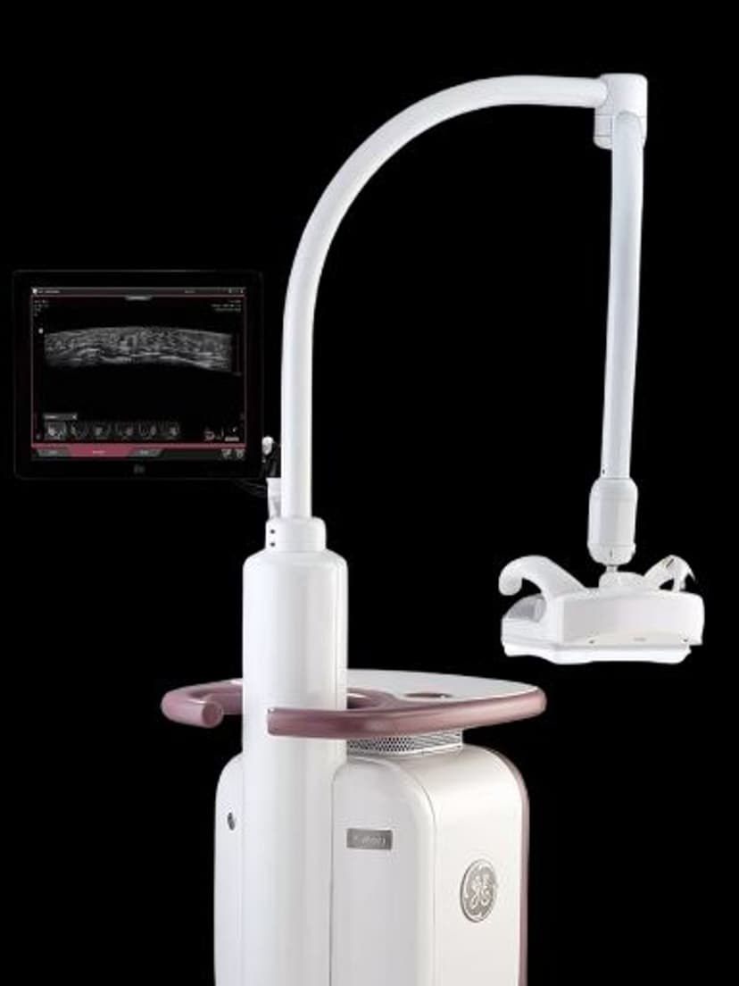
GE Ultrasound Feature
Lesion Segmentation
MeasurementLesion Segmentation is an AI-based measurement tool on GE LOGIQ systems that automatically detects and outlines the boundaries of focal lesions in ultrasound images. Instead of manually tracing lesion margins with a trackball, clinicians activate the tool and the system identifies the lesion edges, calculates volume, and provides dimensional measurements. This automation is particularly valuable in breast, liver, and thyroid imaging, where accurate lesion sizing directly affects clinical staging and treatment planning. Lesion Segmentation also supports serial monitoring by providing consistent boundary definitions across follow-up exams.

Key Benefits
Why Lesion Segmentation matters
Automated boundary detection replaces manual tracing
The AI identifies lesion edges and generates a contour automatically. This eliminates the slow, operator-dependent process of manual trackball tracing, cutting per-lesion measurement time from minutes to seconds.
Reproducible measurements for serial tumor monitoring
Consistent automated boundaries make follow-up comparisons clinically meaningful. When the same algorithm defines the lesion edge at each visit, size changes reflect actual tumor response rather than inter-operator measurement drift.
Multi-lesion detection in a single scan
Lesion Segmentation can identify and outline multiple lesions within the same image frame. Each lesion gets individual volume and dimension calculations, speeding up documentation in patients with multi-focal disease.
Support for BI-RADS and oncology staging documentation
Precise, automated lesion dimensions feed directly into clinical staging workflows. In breast imaging, the measurements support BI-RADS category documentation. In liver and thyroid imaging, the data supports RECIST-based treatment response assessment.
About Lesion Segmentation
Manual lesion tracing in ultrasound is one of the most operator-dependent measurements in diagnostic imaging. Two sonographers tracing the same lesion can produce different boundary outlines, which leads to different volume and dimension calculations. Lesion Segmentation addresses this variability by applying image processing algorithms that detect contrast differences at lesion edges and generate a consistent boundary contour. The system can detect and outline multiple lesions within the same scan, calculating area, perimeter, and estimated volume for each. In oncology workflows, this capability is used for serial tumor monitoring, where small changes in lesion size guide treatment decisions. Consistent automated boundaries make it possible to detect genuine size changes rather than measurement variability between operators. The tool works in real time during live scanning, so clinicians see the segmentation overlay immediately and can verify or adjust the boundary before saving. For breast imaging, Lesion Segmentation supports BI-RADS documentation by providing precise size measurements. In liver imaging, it assists with tracking hepatic lesions across treatment cycles. The feature reduces per-lesion measurement time from minutes of manual tracing to seconds of automated detection.
Availability
Available on these systems

Our Partners


Want Lesion Segmentation in your practice?
Request a quote for a system that includes this feature.