
GE Ultrasound Feature
PDI Quantification
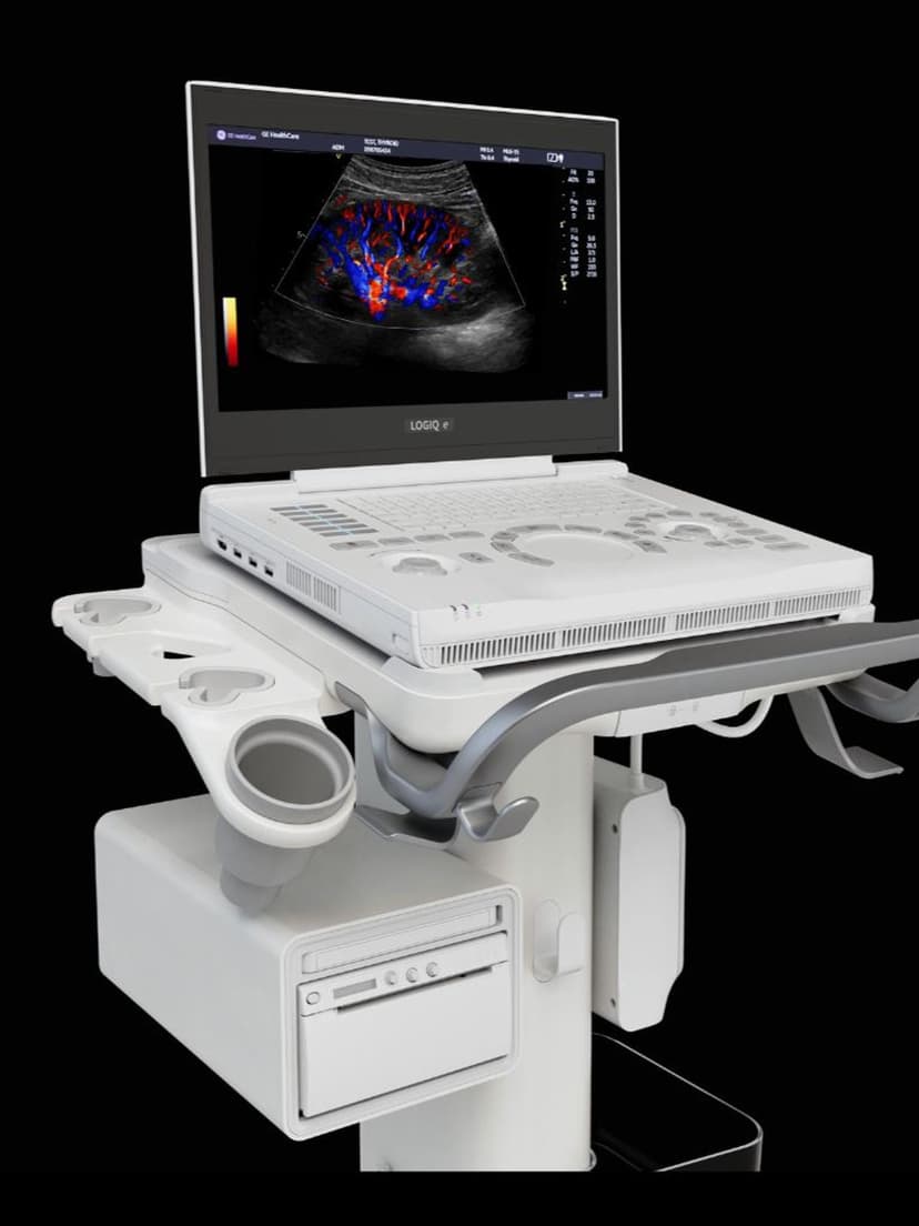
MeasurementPDI Quantification uses Power Doppler Imaging to measure and display blood flow intensity independent of flow direction and velocity angle. Unlike standard color Doppler, Power Doppler detects the amplitude of the Doppler signal rather than frequency shift, making it more sensitive to slow flow and small-vessel perfusion. PDI Quantification adds numerical analysis to this imaging mode, calculating flow area, mean power, and vascularity index within a defined region of interest. This gives clinicians objective, repeatable metrics for assessing organ perfusion, monitoring tumor vascularity, evaluating inflammatory conditions, and tracking treatment response over time.

Key Benefits
Why PDI Quantification matters
Detect blood flow that color Doppler misses
Power Doppler's amplitude-based detection finds slow flow in small vessels and capillary beds where frequency-shift Doppler drops out. This sensitivity matters for assessing organ perfusion, synovial inflammation, and tumor vascularity.
Objective vascularity measurements for serial monitoring
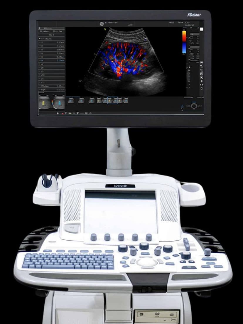
Region-of-interest quantification produces numerical flow metrics that can be compared across exams. Track changes in vascularity index, color pixel area, or mean power over weeks or months to monitor disease progression or treatment response.
Angle-independent flow assessment
Because Power Doppler measures signal amplitude rather than frequency shift, results are not affected by the angle between the transducer and the vessel. This reduces operator-dependent variability and improves measurement reproducibility.
Multi-specialty clinical applications
PDI Quantification supports vascular assessment, rheumatologic synovial scoring, oncologic perfusion monitoring, renal transplant evaluation, and obstetric placental flow analysis. One measurement tool serves multiple clinical workflows.
About PDI Quantification
Standard color Doppler displays flow direction and velocity but loses sensitivity in vessels with slow flow or at steep angles relative to the transducer. Power Doppler addresses this by measuring the integrated power of the Doppler spectrum, which reflects the density of moving blood cells rather than their velocity or direction. The result is a more sensitive flow map that detects perfusion in small vessels, synovial tissue, and organ parenchyma. PDI Quantification extends this capability with region-of-interest analysis tools that calculate vascularity parameters within a defined area. Clinicians can track metrics such as percent vascularity, color pixel area, and mean power values. These quantified measurements enable objective comparisons between serial exams, supporting treatment monitoring in rheumatology (synovial vascularity in inflammatory arthritis), oncology (tumor perfusion changes), transplant medicine (renal graft perfusion), and OB/GYN (placental blood flow assessment). Because Power Doppler is angle-independent, PDI Quantification produces more consistent measurements across operators and scanning positions than velocity-based Doppler indices.


Our Partners


Want PDI Quantification in your practice?
Request a quote for a system that includes this feature.