
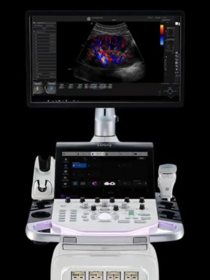
GE Ultrasound Feature
Photo Assistant
WorkflowPhoto Assistant lets clinicians capture external images, such as photos of skin lesions, surgical sites, or anatomical landmarks, using a mobile device or connected camera and embed them directly into the ultrasound exam report. This creates a more complete record by combining ultrasound findings with visual context that B-mode imaging alone cannot capture. The feature is available on GE LOGIQ systems and supports multiple image attachments per exam, adding visual documentation without requiring a separate imaging system or manual file management.

Key Benefits
Why Photo Assistant matters
Unified report with ultrasound and external imagery
External photos are embedded directly into the ultrasound report, creating a single document that combines B-mode findings with visual context. No separate file management or cross-referencing required.
Clearer referral communication
Referring physicians see both the ultrasound findings and the external appearance of the area of interest. This reduces follow-up questions and improves the accuracy of clinical consultations.
Real-time capture during the exam
Photos are taken and attached during the active exam session, not added retroactively. This ensures the images are current and accurately represent the patient's condition at the time of scanning.
Simplified documentation for medico-legal records
Having external photos embedded in the DICOM study creates a timestamped, integrated record. This is valuable for procedures, biopsies, and any exam where visual documentation of the external site strengthens the clinical record.
About Photo Assistant
Clinical documentation often benefits from visual context beyond what ultrasound imaging provides. A dermatology referral may need a photo of the external lesion alongside the ultrasound of the subcutaneous structure. An MSK exam may benefit from a photo showing the patient's range of motion or the exact point of tenderness. Photo Assistant addresses this by allowing clinicians to capture external images during the exam and attach them directly to the DICOM study. The images are embedded in the report alongside the ultrasound data, creating a single unified record that travels with the patient through the medical system. This eliminates the need to manage separate photo files, cross-reference different systems, or rely on written descriptions of visible findings. For practices that handle referrals or multi-disciplinary consultations, Photo Assistant reduces communication gaps by giving the reviewing physician both the ultrasound data and the external visual context in one place.


Our Partners


Want Photo Assistant in your practice?
Request a quote for a system that includes this feature.