
GE Ultrasound Feature
POC Ultrasound
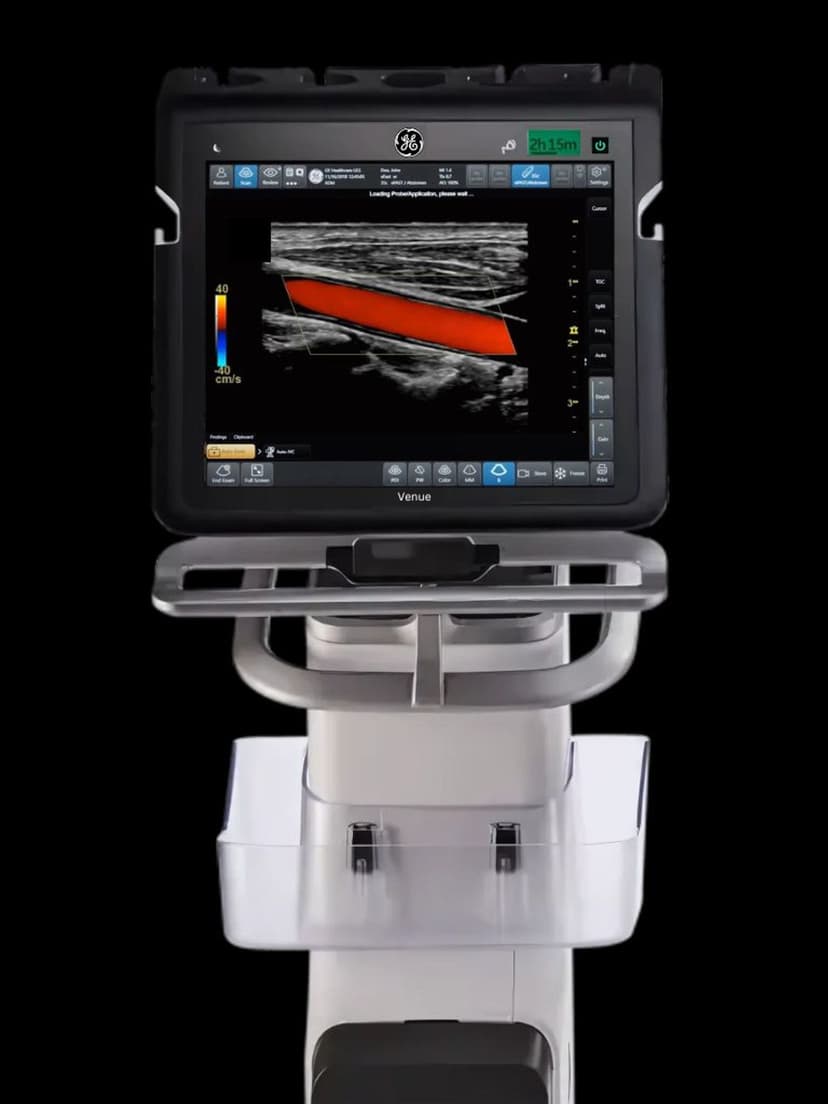
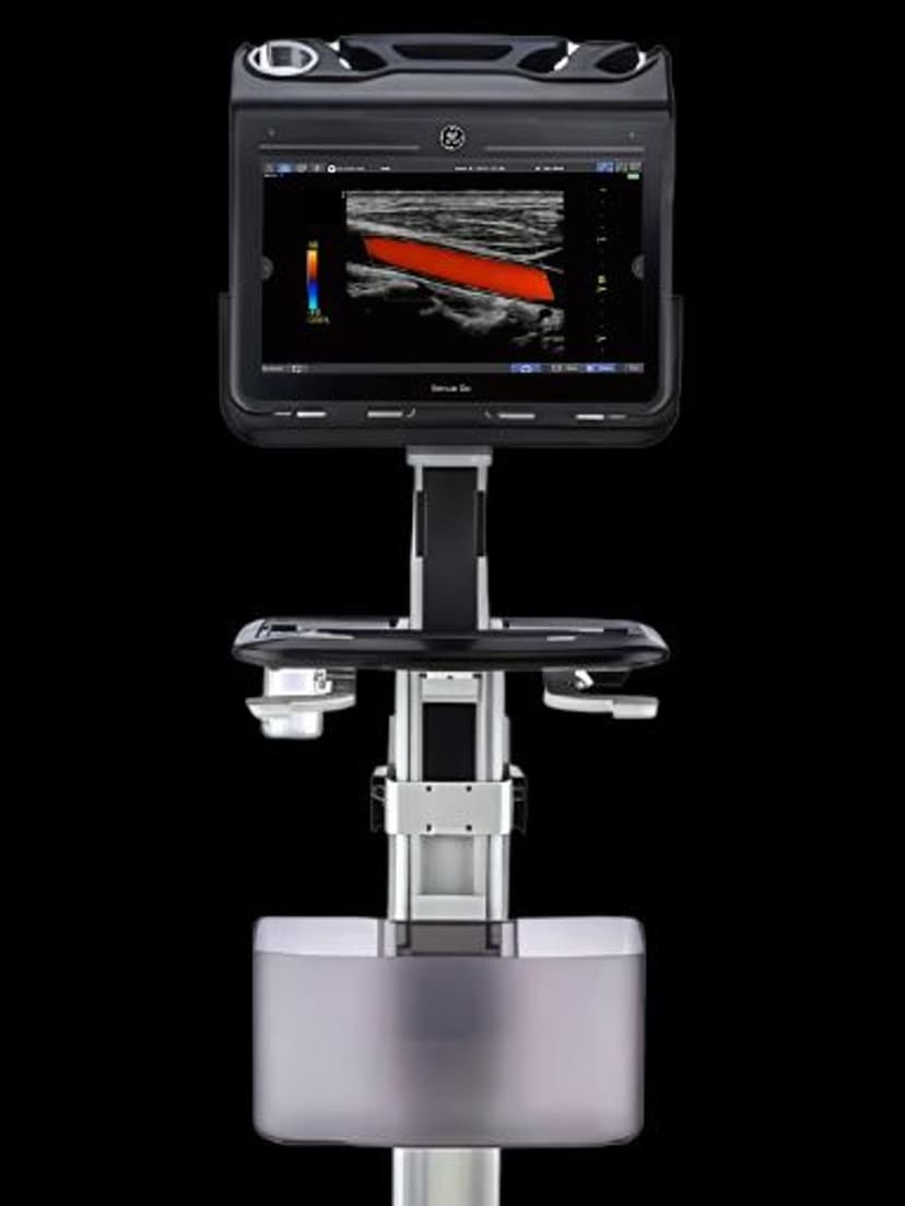
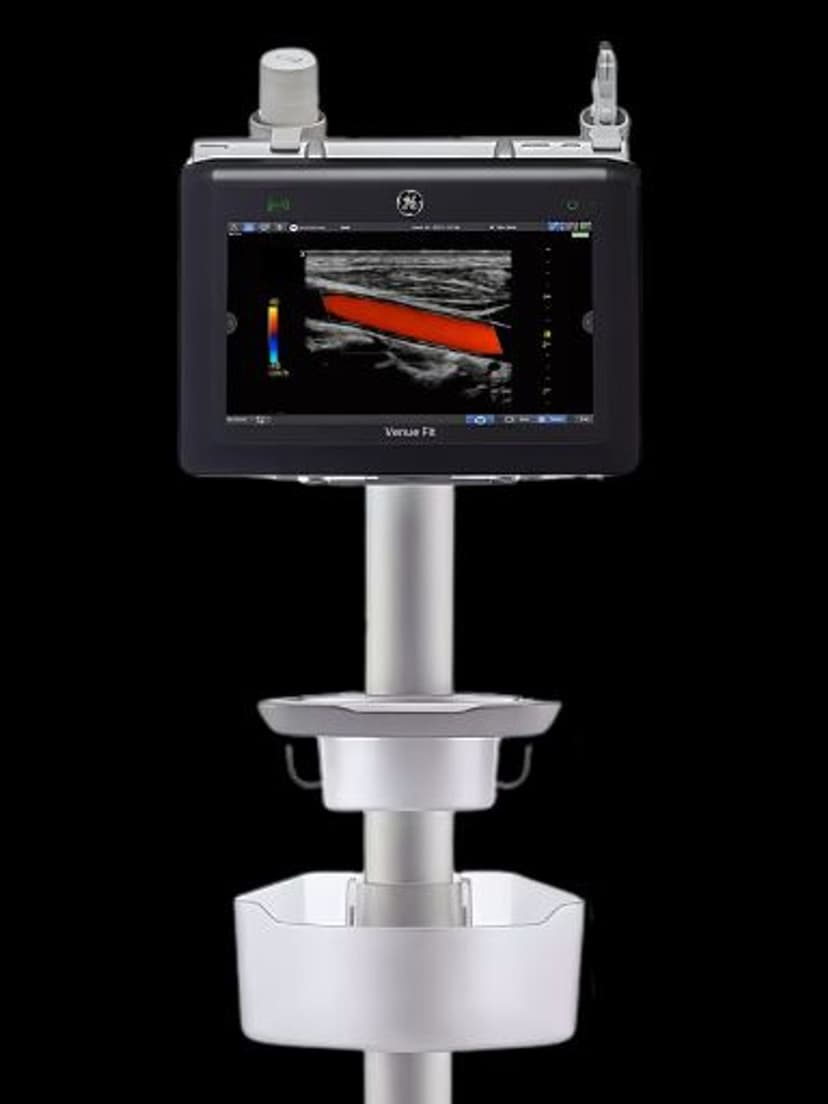
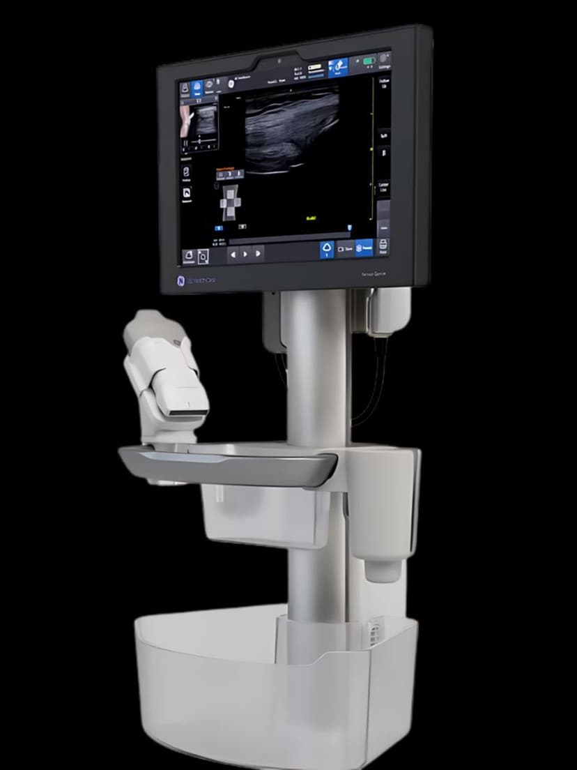
WorkflowPOC Ultrasound is the clinical paradigm the Venue family was engineered around: the clinician brings the ultrasound system to the patient's bedside, scans, and uses the result in the same encounter to guide treatment. On the Venue, Venue Go, Venue Fit, and Venue Sprint, this translates to product decisions that differ from a full diagnostic console: faster boot time, auto-calculation templates for common exam types, and a user interface assuming the operator is a physician or nurse rather than a sonographer. Emergency medicine, critical care, anesthesia, and primary care teams use these systems to answer clinical questions in real time during the patient encounter.

Key Benefits
Why POC Ultrasound matters
Guides interventional procedures in real time
Vascular access, paracentesis, thoracentesis, and nerve blocks succeed on the first pass more often when the operator visualizes the needle during insertion. The Venue family is sized and positioned for one-handed imaging while the clinician performs the procedure with the other hand.
Removes radiology dependency from acute decisions
During off-hours when the radiology reading room is remote or thinly staffed, ED and ICU teams can still answer questions about pericardial effusion, abdominal free fluid, or pulmonary edema without paging for a formal study. The scan stays entirely within the encounter.
Captures billable evidence inside the patient visit
Image clips, measurements, and findings attach to the chart during the encounter itself, supporting the clinical note and the billing code without a separate report handoff. Practices avoid the common gap where a bedside POC scan gets done but never documented.
Supports residency and advanced practice training programs
Emergency medicine and critical care training programs use POC systems as teaching tools alongside the stethoscope. A resident who learned on Venue Go during training rotates into attending roles already fluent in the equipment deployed across the hospital's ED and ICU fleet.
About POC Ultrasound
Traditional diagnostic ultrasound assumes a trained sonographer, a dedicated exam room, a fixed appointment, and a reading radiologist who interprets the study after the fact. POC ultrasound inverts all four assumptions. The Venue systems boot in seconds rather than minutes so that reaching for the probe during an acute event is not gated by system startup. Exam presets for common POC questions — FAST trauma scans, lung sliding for pneumothorax, inferior vena cava collapse for volume status, focused cardiac evaluation — skip directly to the relevant imaging mode and depth without manual parameter tuning. Reports can be completed and stored against the patient's chart during the same encounter, with image capture and annotation built into the single-touch interface. For ED and ICU teams, this shortens the decision loop from 'I need a scan' to 'I have an answer' to minutes rather than the hours it takes to schedule a formal study.
Availability
Available on these systems
Our Partners


Want POC Ultrasound in your practice?
Request a quote for a system that includes this feature.