
GE Ultrasound Feature
Real-time EF
CardiacReal-time EF is a GE Venue feature that continuously calculates left ventricular ejection fraction during live echocardiographic scanning. Unlike conventional EF measurement, which requires the operator to freeze the image, manually trace endocardial borders at end-diastole and end-systole, and then calculate the result, Real-time EF uses automated algorithms to track ventricular wall motion frame by frame and display EF as a continuously updating value. This eliminates post-processing steps and gives clinicians immediate cardiac function data during bedside assessment. The feature is built for point-of-care settings: emergency departments, ICUs, and pre-operative evaluations where knowing EF quickly influences treatment decisions.

Key Benefits
Why Real-time EF matters
EF displayed during live scanning with no freeze-trace-calculate steps
Real-time EF eliminates the manual workflow of freezing, tracing, and calculating. The system tracks ventricular borders continuously and displays EF as a running value. The clinician sees cardiac function data the moment they obtain an apical view.
Immediate cardiac triage in emergency and ICU settings
In the emergency department, knowing whether EF is normal or severely reduced changes the differential diagnosis and treatment plan within minutes. Real-time EF delivers this information during the initial bedside assessment, before a formal cardiology consult.
Accessible to non-cardiologist operators
Manual EF measurement requires trained echo technique. Real-time EF lowers the skill threshold by automating border detection and calculation, allowing emergency physicians, intensivists, and anesthesiologists to obtain EF data without dedicated echo training.
Serial EF tracking for monitoring treatment response
For ICU patients on inotropic support or undergoing fluid resuscitation, tracking EF over hours helps guide therapy adjustments. Real-time EF makes repeat measurements fast enough to be practical for serial monitoring rather than single-point-in-time assessment.
About Real-time EF
Ejection fraction is the single most important number in cardiac function assessment. A normal EF is 55-70%, and values below 40% indicate significant systolic dysfunction that changes patient management. In traditional echocardiography, EF measurement requires acquiring apical four-chamber and two-chamber views, freezing end-diastolic and end-systolic frames, manually tracing the endocardial border in each frame, and applying Simpson's biplane method. This process takes 2-5 minutes and requires echo training. Real-time EF on GE Venue automates this workflow by applying border detection algorithms to the live image stream. The system identifies the left ventricular cavity in each frame, tracks wall motion across the cardiac cycle, and calculates EF continuously. The value updates with each heartbeat and displays on screen as a running measurement. For point-of-care clinicians who need to know whether cardiac function is normal, moderately reduced, or severely impaired, Real-time EF provides that answer in seconds rather than minutes. The feature does not replace a full echocardiographic study but fills the clinical gap between having no cardiac function data and waiting for a formal echo order to be completed.
Availability
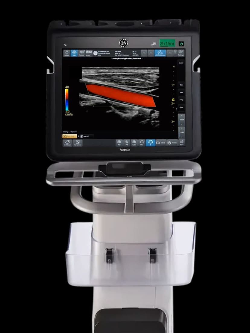
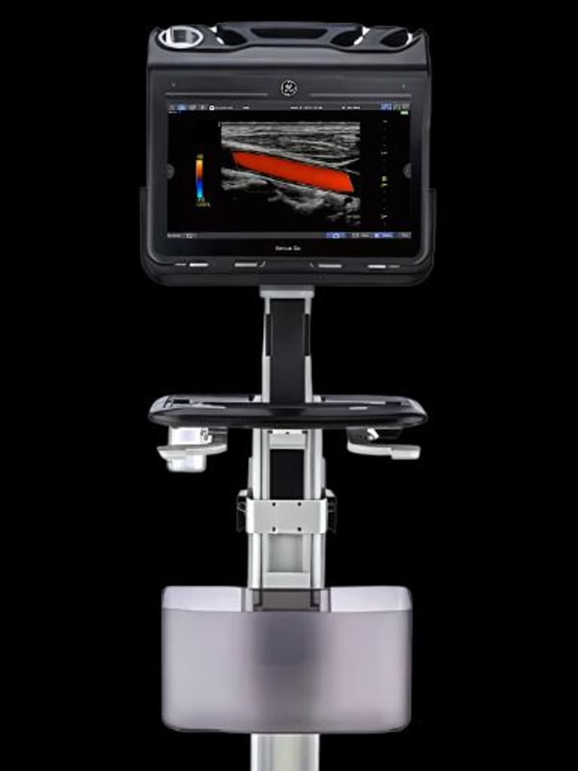
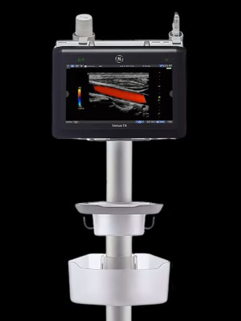
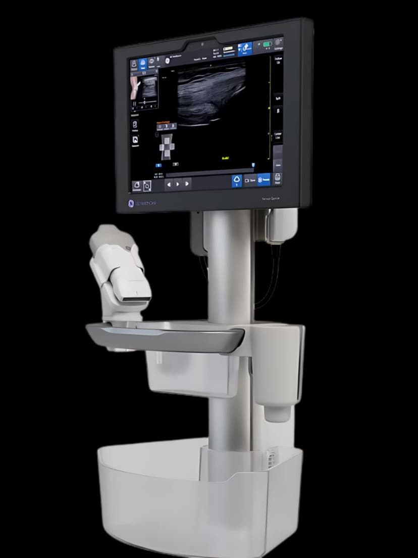
Available on these systems
Our Partners


Want Real-time EF in your practice?
Request a quote for a system that includes this feature.