
GE Ultrasound Feature
Shear Wave
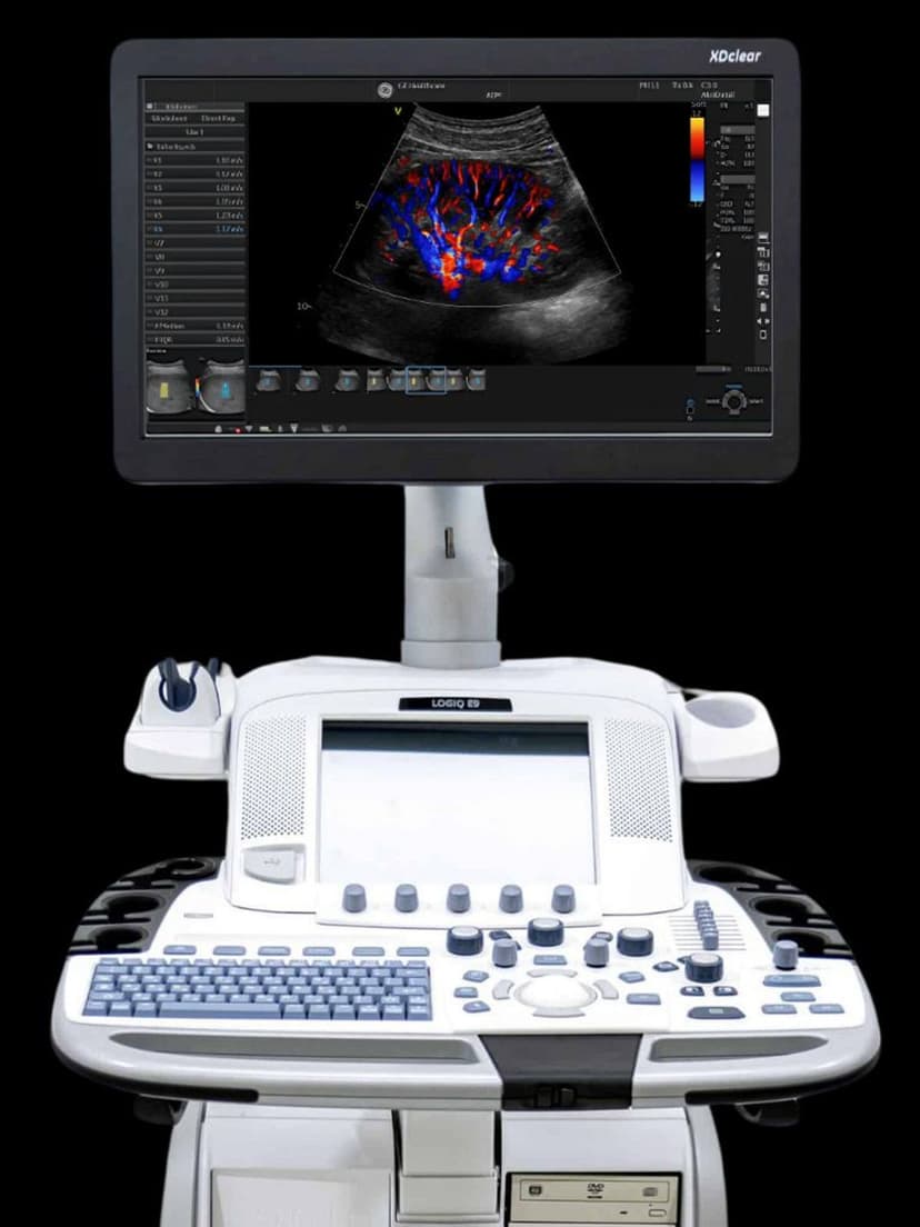
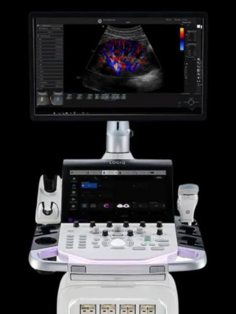
MeasurementShear Wave Elastography on GE LOGIQ systems measures tissue stiffness by generating acoustic radiation force impulse (ARFI) push pulses that create shear waves within the tissue. The system tracks shear wave propagation speed, which correlates directly with tissue stiffness: stiffer tissue transmits shear waves faster. Results display as quantitative values in kilopascals (kPa) or meters per second (m/s), overlaid on a color-coded elasticity map. This gives radiologists, hepatologists, and general imagers an objective, reproducible metric for tissue characterization that replaces subjective palpation and, in many liver fibrosis cases, reduces the need for biopsy.

Key Benefits
Why Shear Wave matters
Quantitative tissue stiffness in kPa or m/s
Shear Wave Elastography replaces subjective assessments with numerical stiffness values. These measurements are reproducible across operators and visits, enabling reliable longitudinal tracking of conditions like liver fibrosis progression or treatment response.
Reduced need for liver biopsy in fibrosis staging
Shear wave stiffness values map directly to fibrosis staging scales recognized by clinical guidelines. For many patients with chronic liver disease, elastography provides sufficient staging information to defer percutaneous biopsy, reducing patient risk and procedure cost.
Color-coded elasticity maps for spatial assessment
A real-time color overlay displays the distribution of tissue stiffness across the measurement region. This spatial mapping reveals focal areas of increased stiffness that may not be apparent on standard B-mode imaging, supporting targeted follow-up or biopsy guidance.
Multi-organ application from liver to breast to MSK
The same shear wave technology applies across clinical departments. Liver fibrosis staging, breast lesion characterization, thyroid nodule assessment, and musculoskeletal tendon evaluation all use tissue stiffness data, making the feature valuable across multiple service lines within a practice.
About Shear Wave
GE's Shear Wave Elastography uses ARFI-based push pulses from the imaging transducer to generate shear waves perpendicular to the ultrasound beam. Tracking pulses then measure the lateral propagation speed of these shear waves through the tissue. The measurement region of interest can be positioned at various depths, and the system calculates mean stiffness values along with confidence metrics. In liver applications, shear wave speed values map to established fibrosis staging scales (F0 through F4), enabling radiologists to stage liver fibrosis without percutaneous biopsy in many patients. Clinical guidelines from AASLD and EASL recognize elastography as an acceptable first-line assessment for chronic hepatitis B and C fibrosis evaluation. Beyond the liver, Shear Wave Elastography applies to breast lesion characterization, where stiffness values help differentiate benign from suspicious masses and may reduce unnecessary biopsy. Thyroid nodule assessment and musculoskeletal applications for tendon and muscle evaluation are also supported. The color-coded stiffness map provides spatial visualization of elasticity distribution within the tissue, allowing clinicians to identify focal areas of increased stiffness within an otherwise normal-appearing region on B-mode imaging.


Our Partners


Want Shear Wave in your practice?
Request a quote for a system that includes this feature.