
GE Ultrasound Feature
Sono Render
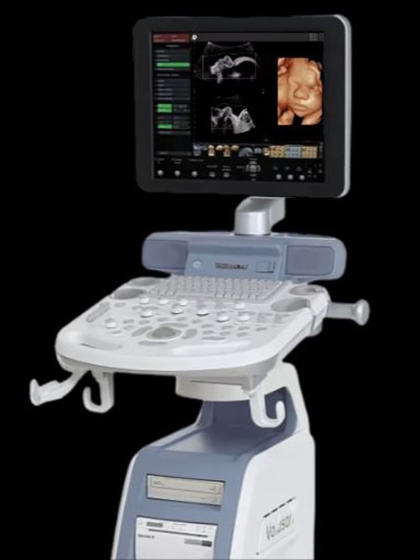
OB/GYNSono Render is GE's 3D/4D rendering toolset on Voluson ultrasound systems, designed to produce detailed surface-rendered images from volume data. After the clinician acquires a 3D or 4D volume, Sono Render processes the dataset to display realistic surface images of fetal anatomy, organ structures, or gynecological findings. The rendering engine automatically adjusts parameters based on the exam type and can be refined manually. OB/GYN sonographers use Sono Render daily for fetal anatomy assessments, parent bonding images, and documentation of structural findings.

Key Benefits
Why Sono Render matters
Detailed surface rendering of fetal anatomy
Sono Render converts raw 3D volume data into recognizable surface images showing fetal facial features, limbs, and body contours. These rendered images support diagnostic assessment of surface abnormalities and provide parents with clear images of their developing baby.
Real-time 4D rendering during live scanning
In 4D mode, Sono Render processes and displays volume data continuously, showing fetal movement in real time. Clinicians can observe swallowing, yawning, and limb movement as it happens, adding functional assessment capabilities to the structural anatomy scan.
Multiple visualization modes for different clinical needs
Surface rendering shows external anatomy, transparent mode reveals internal structures, and maximum intensity projection maps vascularity. One volume acquisition can produce multiple diagnostic views without rescanning the patient.
Preset profiles reduce manual adjustment time
Sono Render includes optimization presets calibrated for common OB scenarios like fetal face imaging, limb evaluation, and spine assessment. Sonographers spend less time adjusting render parameters manually and more time on clinical assessment and patient interaction.
About Sono Render
Sono Render processes raw 3D volume data through a multi-step rendering pipeline. The system first identifies the region of interest within the acquired volume, then applies surface detection algorithms to distinguish tissue boundaries from surrounding fluid or artifact. A combination of threshold adjustment, smoothing, and lighting simulation produces the final rendered image. In obstetrics, this means converting a raw 3D dataset of the fetal head into a recognizable face with realistic surface features. The rendering engine supports multiple visualization modes including surface rendering, transparent rendering for internal structures, and maximum intensity projection for vascular mapping. Sono Render works with both static 3D acquisitions and live 4D scanning. During 4D mode, the system renders volumes in real time, allowing clinicians and parents to see fetal movement as it happens. The quality of the rendered output depends on several factors: amniotic fluid volume, fetal position, maternal body habitus, and probe frequency. Sono Render includes preset optimization profiles for common OB scenarios, reducing the manual adjustment needed to produce a clear result. For practices that offer keepsake imaging services alongside diagnostic scanning, Sono Render provides the visual quality that parents expect.
Availability
Available on these systems
Our Partners


Want Sono Render in your practice?
Request a quote for a system that includes this feature.