
GE Ultrasound Feature
SonoCNS
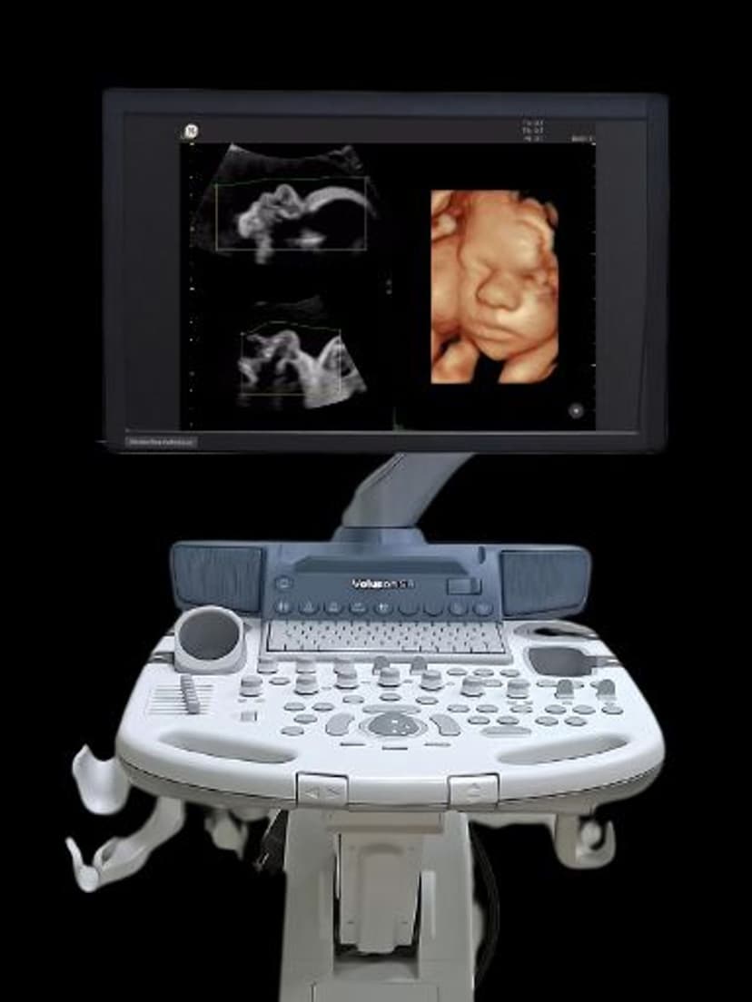
OB/GYNSonoCNS automates the measurement of fetal central nervous system structures on GE Voluson ultrasound systems. From a single 3D volume acquisition of the fetal head, SonoCNS identifies standard anatomical planes and measures structures including the lateral ventricles, cerebellum, cisterna magna, cavum septi pellucidi, and biparietal diameter. The feature replaces what would otherwise require manual navigation through multiple 2D planes and individual caliper placements. Maternal-fetal medicine specialists and OB/GYN sonographers use SonoCNS to perform faster, more standardized neurosonography assessments during second- and third-trimester anatomy scans.

Key Benefits
Why SonoCNS matters
Nine CNS planes from a single 3D volume
Instead of manually navigating to each anatomical plane and placing calipers individually, SonoCNS extracts up to nine standard views from one volume acquisition. This turns a multi-step measurement workflow into a semi-automated process that takes a fraction of the time.
Standardized neurosonography across operators
Fetal brain measurement is one of the most variable components of OB ultrasound. SonoCNS applies the same algorithmic approach regardless of who performs the scan, reducing inter-operator variability and improving consistency for multi-site practices and high-volume OB clinics.
Early detection of ventriculomegaly and CNS anomalies
SonoCNS measures lateral ventricle width with reference to gestational age norms. Values exceeding 10mm are automatically flagged, supporting early identification of ventriculomegaly and prompting timely referral to maternal-fetal medicine specialists.
Summary screen with reference ranges for quick review
All CNS measurements display on a single results screen alongside gestational age-matched reference ranges. Clinicians can review the entire fetal brain assessment at a glance and quickly identify any measurement that falls outside normal limits, rather than scrolling through individual image frames.
About SonoCNS
Fetal neurosonography is one of the most operator-dependent components of the obstetric anatomy scan. Identifying the correct axial, coronal, and sagittal planes through the fetal brain requires experience, and manual measurements across multiple planes are time-consuming and prone to inter-operator variability. SonoCNS addresses both problems by working from a 3D volume dataset. After the clinician acquires a 3D volume of the fetal head, the software automatically identifies up to nine standard CNS planes and places measurement calipers on key structures. The system measures the atrial width of the lateral ventricles (the primary marker for ventriculomegaly), transcerebellar diameter, cisterna magna depth, and other biometric parameters. All measurements display on a single summary screen with reference ranges, making it easy to identify values outside normal limits. SonoCNS is particularly valuable for detecting ventriculomegaly, posterior fossa abnormalities, and midline structural anomalies. The standardized approach means results are consistent whether the scan is performed by a senior perinatologist or a general OB sonographer, which is critical for practices that rely on multiple operators across locations.
Availability
Available on these systems
Our Partners


Want SonoCNS in your practice?
Request a quote for a system that includes this feature.