
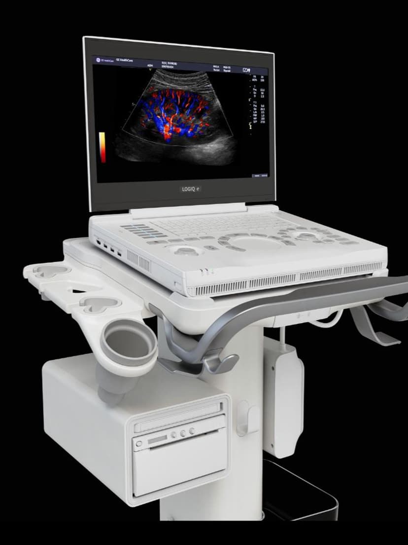
GE Ultrasound Feature
Split Screen
WorkflowSplit Screen divides the ultrasound display into two or more panels so clinicians can view different imaging modes simultaneously during a live exam. One panel might show a real-time B-mode image while the adjacent panel displays Color Doppler, PW Doppler, or a previously saved reference scan. This eliminates the need to toggle between modes, which saves time and reduces the risk of losing a scanning window. Cardiac exams benefit when comparing apical views across different Doppler measurements. Vascular studies gain efficiency by placing B-mode anatomy next to spectral Doppler waveforms in a single glance. General imaging workflows use it to compare a current scan against a prior study stored on the system, supporting interval change assessment without leaving the exam screen.

Key Benefits
Why Split Screen matters
Compare imaging modes without toggling views
Split Screen places B-mode, Doppler, and other modalities side by side on a single display. Clinicians see both views at once rather than switching back and forth, which preserves the scanning window and reduces the chance of losing anatomical orientation.
Faster duplex and multi-mode exams
Vascular and cardiac exams that require paired views, such as B-mode anatomy alongside spectral Doppler, can be completed more quickly. The sonographer captures both perspectives in a single screen state instead of separate acquisition steps.
Side-by-side comparison with prior studies
Clinicians can pull a stored reference image into one panel while scanning live in the other. This supports interval change assessment during follow-up exams, such as tracking lesion size or monitoring treatment response over time.
Composite screenshots for cleaner documentation
Paired images captured in Split Screen mode save as a single frame in the patient record. This produces cleaner, more informative exam documentation compared to saving each view separately, and it reduces the number of individual images archived per study.
About Split Screen
Split Screen is a display management feature available on GE ultrasound systems that partitions the monitor into separate viewing areas. Each panel operates independently, allowing clinicians to assign different imaging modes, freeze states, or stored images to each section. A sonographer performing a carotid duplex exam can display a longitudinal B-mode view of the carotid bifurcation on one side while capturing PW Doppler spectral waveforms on the other. Cardiac sonographers use it to place a parasternal long-axis view beside an apical four-chamber view for simultaneous assessment. The layout is adjustable based on clinical need, with options for left-right or top-bottom configurations depending on the system. Because both panels update from the same transducer connection, there is no delay in switching context. The feature reduces physical interaction with the keyboard by removing the need to cycle between full-screen modes, and it supports faster reporting by presenting paired images that can be saved as a single composite screenshot for the patient record.
Availability
Available on these systems
Our Partners


Want Split Screen in your practice?
Request a quote for a system that includes this feature.