
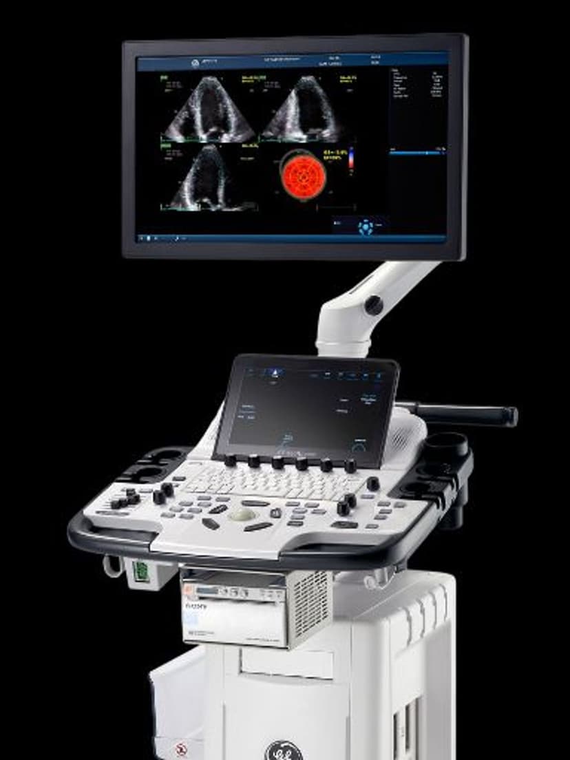
GE Ultrasound Feature
Strain Elastography
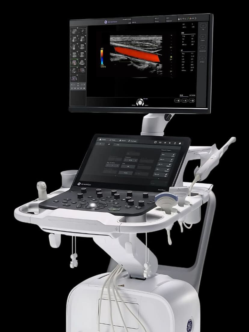
MeasurementStrain Elastography measures how tissue deforms under applied compression, producing a color-coded elastogram overlaid on the B-mode image. The clinician applies gentle rhythmic pressure with the probe, and the system compares tissue displacement frame-to-frame to calculate relative stiffness. Stiffer tissues (suspicious masses, fibrotic areas) show less deformation than surrounding normal tissue, appearing as distinct color regions on the elastogram. This additional stiffness data helps clinicians characterize breast lesions, thyroid nodules, and musculoskeletal abnormalities without biopsy. In breast imaging, strain elastography improves lesion specificity by adding an elasticity dimension to B-mode morphology and Doppler flow findings.

Key Benefits
Why Strain Elastography matters
Improved breast lesion characterization without biopsy
Strain elastography adds tissue stiffness data to standard B-mode and Doppler findings. For BI-RADS 3 and 4a lesions, the elasticity information can improve specificity and help reduce the rate of unnecessary biopsies of benign masses.
Quantitative stiffness ratios for objective assessment
The strain ratio and E/B ratio provide numerical values that support clinical decision-making beyond subjective visual interpretation. These quantified measurements can be documented, compared over time, and communicated clearly in reports.
No additional hardware required
Strain elastography uses the existing ultrasound probe and manual compression. There is no need for specialized shear wave hardware or push-pulse transducers, making it available on a broader range of GE systems at a lower cost.
Multi-organ application from breast to MSK
The same strain elastography tool applies to breast lesions, thyroid nodules, and musculoskeletal soft tissue assessment. Practices performing imaging across these specialties get diagnostic value from a single feature across multiple exam types.
About Strain Elastography
Strain elastography compares ultrasound frames acquired during gentle probe compression to map how much tissue deforms relative to adjacent tissue. Softer tissue compresses more; stiffer tissue compresses less. The system converts that relative deformation into a color map over the B-mode image and can provide quantification tools such as strain ratio or elastogram-to-B-mode (E/B) comparison. Because the measurement is relative rather than absolute, strain elastography is more operator-dependent than shear wave techniques, but it works well for superficial structures where controlled compression is feasible. In breast imaging, it adds stiffness context to BI-RADS assessment, helping separate softer benign-appearing lesions from stiffer suspicious ones. Three quantification methods are available: strain ratio compares target to reference tissue stiffness, E/B ratio compares lesion dimensions on elastogram versus B-mode, and the qualitative color scale supports rapid screening. In thyroid and musculoskeletal exams, it adds information about nodule firmness or tendon degeneration that may not be obvious on grayscale imaging alone. Unlike shear wave elastography, which provides absolute stiffness in kilopascals, strain elastography measures relative stiffness within the field of view.
Availability
Available on these systems

Our Partners


Want Strain Elastography in your practice?
Request a quote for a system that includes this feature.