
GE Ultrasound Feature
Venue View
Imaging TechnologyVenue View is a panoramic imaging feature on GE Venue-series ultrasound systems that extends the field of view beyond a single transducer footprint. The system stitches consecutive image frames together in real time as the clinician sweeps the probe across the anatomy, producing one continuous wide-field image. This is especially useful for visualizing structures that exceed the probe's native field of view, such as long bones, large abdominal organs, thyroid beds, or musculoskeletal structures. Venue View eliminates the need to mentally reconstruct anatomy from multiple separate images, giving clinicians a single reference frame for measurement and documentation.

Key Benefits
Why Venue View matters
Full anatomical context in a single image
Captures structures larger than the probe footprint in one continuous sweep. Clinicians no longer need to mentally piece together separate images when assessing long bones, tendons, or large organ spans.
More accurate measurements of extended structures
Measuring a structure that spans multiple standard frames introduces alignment error. Venue View provides a single unified image for end-to-end measurement, improving measurement precision for long anatomical structures.
Faster documentation with fewer saved images
One panoramic capture replaces several individual frames. This reduces image storage volume and speeds up reporting, since clinicians document the finding once rather than cross-referencing multiple clips.
Wider context for interventional needle tracking
During procedures such as biopsies or catheter placements, Venue View provides an extended field that shows both the needle entry point and the target in a single continuous display. This broader perspective supports safer needle guidance in point-of-care settings.
About Venue View
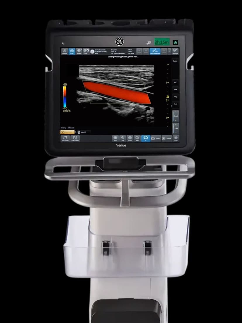
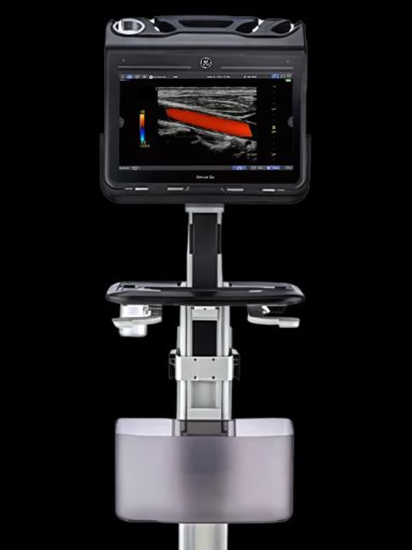
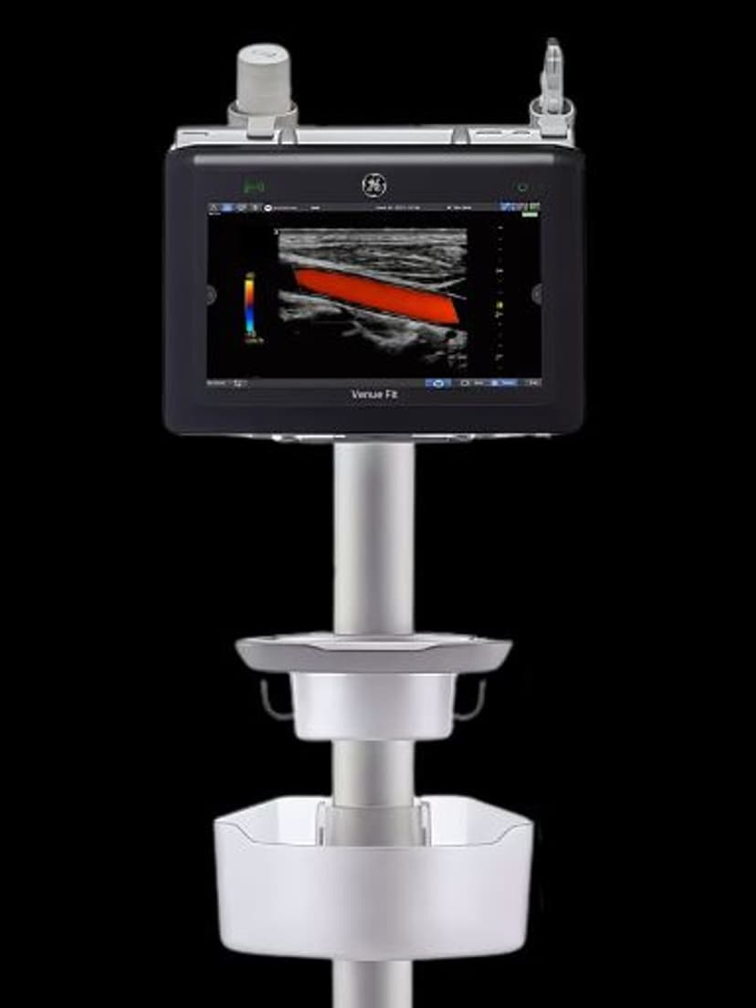
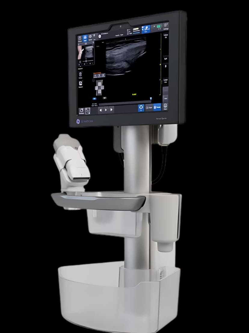
Venue View uses real-time image registration and stitching algorithms to combine sequential B-mode frames into a single panoramic display. As the clinician moves the probe along the anatomy, the system tracks frame-to-frame motion and aligns each new frame with the previous ones, building a continuous extended image. Unlike static field-of-view limitations inherent to any single transducer, Venue View lets clinicians capture the full extent of a structure in one sweep. This is particularly valuable in musculoskeletal imaging, where tendons, ligaments, and long bones often extend well beyond a standard linear probe footprint. In abdominal imaging, Venue View provides a wider context for measuring organ dimensions or tracking the extent of pathology. The panoramic image can be saved and measured directly on the system. For interventional applications, the extended view helps track needle paths across a wider anatomical field during procedures like biopsies. Venue View requires no additional hardware and works with the standard probes available on Venue Sprint and Venue Fit systems.
Availability
Available on these systems
Our Partners


Want Venue View in your practice?
Request a quote for a system that includes this feature.