
GE Ultrasound Feature
Virtual Convex
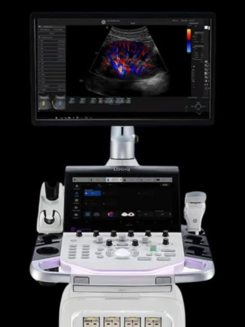
Imaging TechnologyVirtual Convex digitally expands the field of view of a linear transducer beyond its physical aperture, creating a trapezoidal or curved display similar to what a convex probe would produce. The software steers the ultrasound beam beyond the edges of the linear array, widening the viewing angle while maintaining the high-frequency resolution that linear probes provide. This eliminates the need to switch to a lower-frequency convex probe when a broader view is needed during vascular, musculoskeletal, or emergency imaging, saving time and maintaining image quality for superficial structures.

Key Benefits
Why Virtual Convex matters
Wider field of view without changing probes
Virtual Convex expands the viewing angle of a linear transducer beyond its physical aperture. Clinicians see more anatomy in a single image without stopping the exam to switch to a convex probe, which saves time and maintains workflow momentum.
High-frequency resolution preserved across the wider view
Unlike switching to a convex probe that operates at lower frequencies, Virtual Convex maintains the linear probe's high-frequency resolution in the center of the image. Superficial structures remain sharply defined even as the field of view expands.
Better anatomical context for targeted exams
Seeing the vessel, nerve, or structure of interest within its broader anatomical surroundings improves orientation and diagnostic confidence. This is particularly useful during vascular mapping, MSK assessment, and interventional procedures where spatial relationships matter.
Useful in emergency settings where speed matters
In trauma and emergency departments, operators often start with a linear probe for focused assessment. Virtual Convex lets them survey a wider area without the delay of probe changes, supporting faster clinical decision-making when time is critical.
About Virtual Convex
Linear probes produce a rectangular field of view limited to the physical width of the transducer footprint, typically 38-50 mm for standard linear arrays. While this is adequate for focused superficial imaging, many clinical situations require a wider anatomical perspective. Traditionally, the operator would switch to a convex probe to get a broader view, but convex probes operate at lower frequencies and sacrifice the fine resolution needed for superficial detail. Virtual Convex solves this tradeoff by electronically steering the beam at the edges of the linear array to create a wider field of view. The center of the image retains the full linear resolution while the lateral edges extend outward at an angle, producing a trapezoidal display. This gives clinicians the context of surrounding anatomy without losing detail in the region of interest. In vascular imaging, Virtual Convex helps visualize the full extent of a vessel and its relationship to adjacent structures. In MSK imaging, it provides a broader view of muscle groups and joint spaces. The feature adjusts in real time, and the operator can toggle it on or off during scanning without interrupting the exam.


Our Partners


Want Virtual Convex in your practice?
Request a quote for a system that includes this feature.