
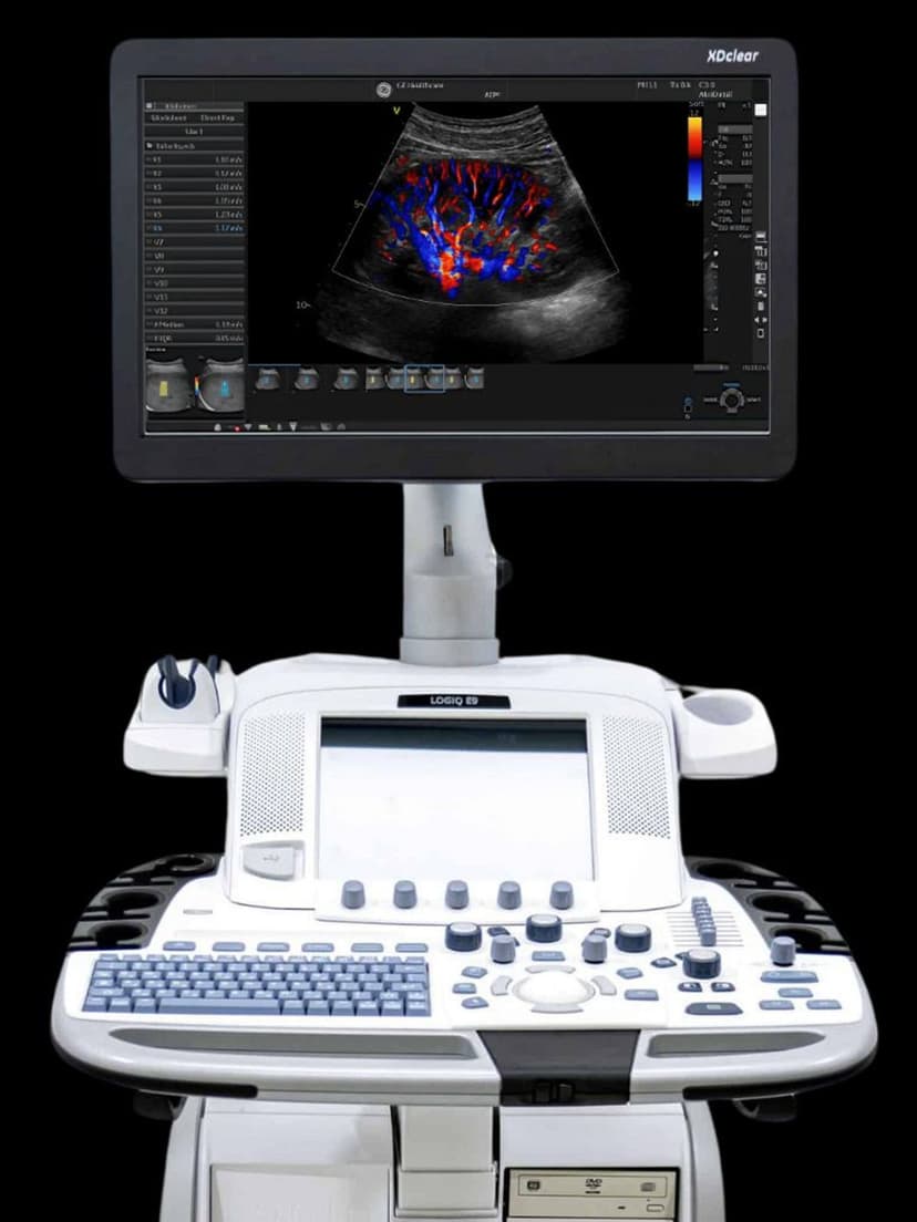
GE Ultrasound Feature
Volume Navigation
InterventionalVolume Navigation overlays live ultrasound imaging onto previously acquired CT or MRI 3D datasets, giving clinicians a synchronized multimodal view during interventional procedures. The system registers and tracks the ultrasound probe position relative to the stored volume, so needle paths and anatomical landmarks from cross-sectional imaging stay visible in real time. This is particularly valuable for liver biopsies and ablations where ultrasound alone may not clearly show the target lesion. Radiologists and interventionalists use Volume Navigation to reach targets identified on CT or MRI with the flexibility and real-time feedback of ultrasound guidance.

Key Benefits
Why Volume Navigation matters
Target lesions invisible on ultrasound alone
Some lesions are clearly visible on CT or MRI but blend into surrounding tissue on B-mode ultrasound. Volume Navigation fuses both modalities so you can use CT/MRI conspicuity while maintaining the real-time flexibility of ultrasound-guided needle placement.
Real-time probe tracking across modalities
The system tracks ultrasound probe position and continuously updates the corresponding CT or MRI plane. This live synchronization eliminates the mental mapping between stored cross-sectional images and live ultrasound, reducing procedural uncertainty.
Improved accuracy for liver biopsies and ablations
Hepatology and interventional radiology teams use Volume Navigation to guide biopsies and thermal ablations with higher confidence. The fused view helps confirm needle position relative to the target and adjacent critical structures before tissue sampling or energy delivery.
Fewer repeat imaging studies and procedure deferrals
When a target cannot be found on ultrasound alone, the procedure is often deferred and a repeat CT is ordered. Volume Navigation reduces these deferrals by making previously identified lesions visible through fusion, saving time for both the patient and the department.
About Volume Navigation
Volume Navigation works by co-registering a live ultrasound image with a previously acquired 3D dataset, typically from CT or MRI. The clinician selects anatomical reference points visible on both modalities, and the system calculates a spatial transformation that locks the two datasets together. As the ultrasound probe moves, the corresponding CT or MRI slice updates in real time on a side-by-side or overlay display. This fusion approach is especially useful for targeting lesions that are conspicuous on CT or MRI but difficult to see on B-mode ultrasound, such as small hepatocellular carcinomas or isoechoic liver metastases. The technology supports freehand needle tracking, allowing the operator to visualize the predicted needle path relative to both the live ultrasound and the reference volume. Clinical studies have shown improved targeting accuracy for liver interventions when fusion guidance is used compared to ultrasound alone, particularly for lesions smaller than 2 cm.
Availability
Available on these systems


Our Partners


Want Volume Navigation in your practice?
Request a quote for a system that includes this feature.