
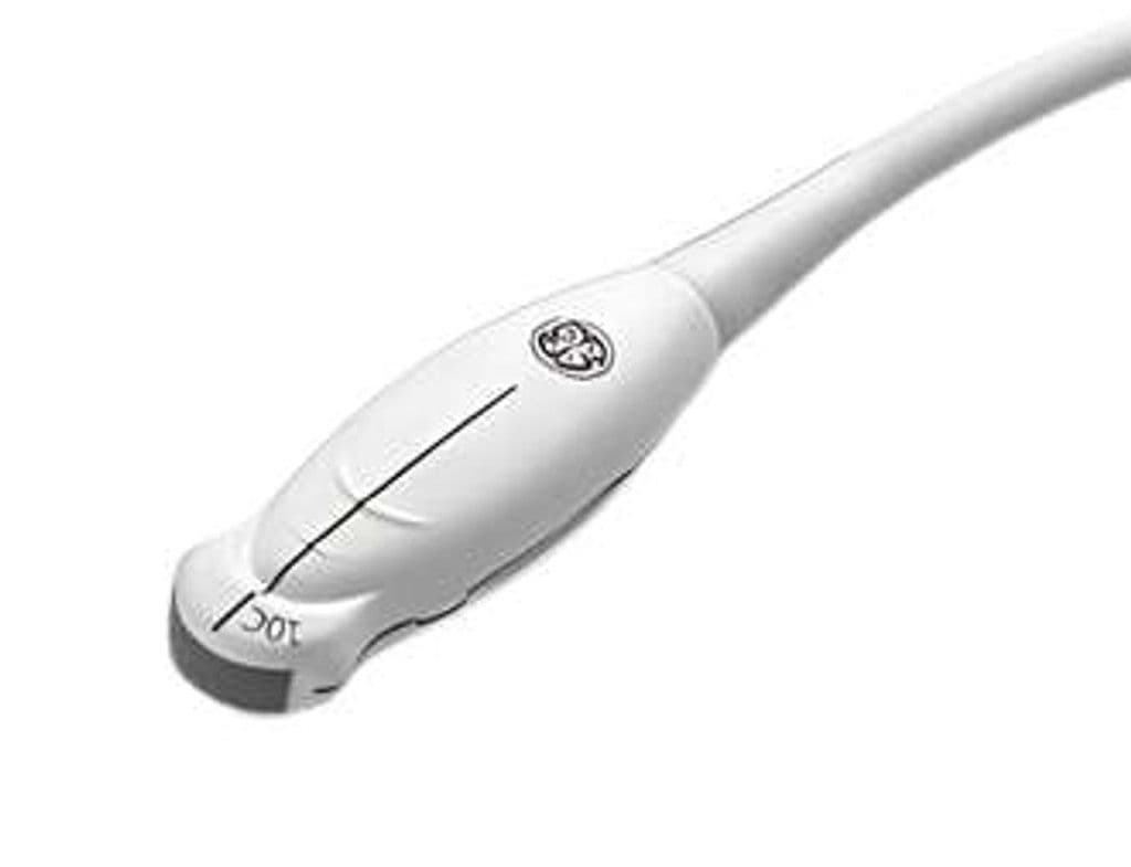
GE 10C-D
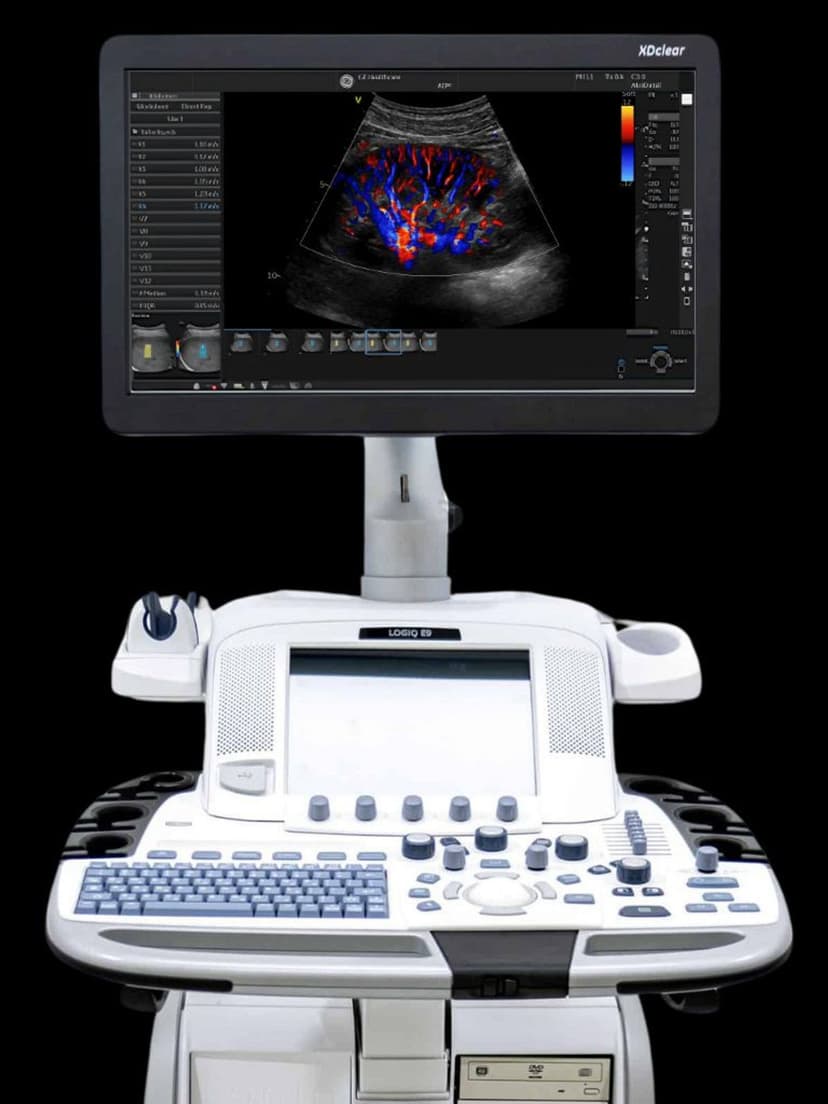
The 10C-D is a microconvex transducer with a compact curved footprint that seats on the neonatal fontanelle and fits between pediatric ribs for abdominal and vascular assessment. The 70-degree sector and 4–12 MHz bandwidth cover superficial cranial structures at high resolution and reach abdominal organs up to 12 cm deep. On LOGIQ P9, P10, and E9 systems, the microconvex geometry provides a wider near-field view than linear probes while maintaining a small contact area.
Compatible Systems

Specifications
Broad bandwidth covers neonatal cranial imaging at high frequency through deeper pediatric abdominal structures.
Wide sector captures both lateral ventricles in a single coronal image through the fontanelle.
Reaches pediatric abdominal organs while maintaining resolution for superficial cranial structures.
Compact curved aperture fits the neonatal fontanelle and pediatric intercostal spaces.
Full diagnostic mode set for tissue morphology and pediatric vascular hemodynamics.
D-Pin interface connects to GE LOGIQ general imaging systems.
Applications
Neonatal Cranial Imaging
The microconvex footprint seats directly on the anterior fontanelle for coronal and sagittal views of the neonatal brain. At 8–12 MHz, the 10C-D differentiates periventricular white matter from gray matter for detection of intraventricular hemorrhage, periventricular leukomalacia, and hydrocephalus. The 70-degree sector angle captures both lateral ventricles in a single coronal image, reducing scan time in the NICU.
Pediatric Abdominal Imaging
The 4 MHz low end penetrates to 12 cm for liver, kidney, and spleen assessment in infants and young children. The compact curved footprint maintains acoustic contact between small ribs and around bowel gas better than larger convex probes. Color Doppler evaluates renal perfusion, portal flow, and hepatic vasculature in patients from preterm neonates through toddlers.
Pediatric Vascular Assessment
The microconvex geometry provides a wide field of view near the skin surface for mapping superficial vessels in small patients. PW Doppler measures flow velocities in renal arteries, mesenteric vessels, and the circle of Willis through the fontanelle. The high-frequency end resolves vessel wall detail in arteries as small as 2–3 mm in diameter.
Our Partners


Need this probe for your system?
Request a quote with pricing, compatibility verification, and delivery timeline.