
GE 10T-D
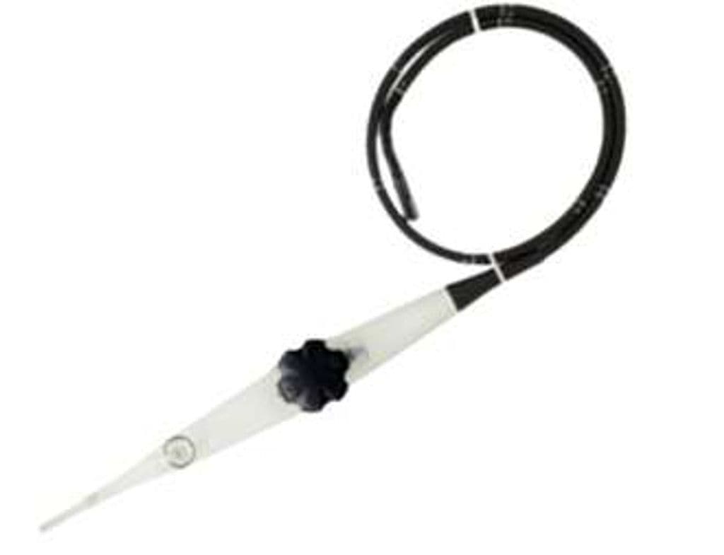
The 10T-D is a transesophageal echocardiography probe with a phased array element behind a miniaturized tip that passes through the esophagus to image the heart from centimeters away. The 3.3–10 MHz bandwidth covers both adult and larger pediatric patients, with high-frequency operation resolving valve leaflet morphology and low-frequency penetration reaching the descending aorta. The slim shaft profile reduces patient discomfort during prolonged intraoperative and ICU monitoring cases.
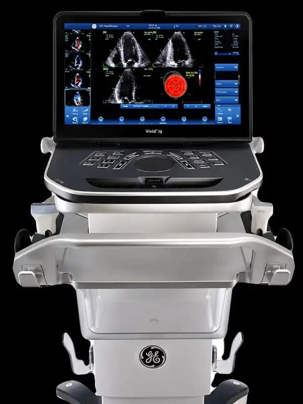
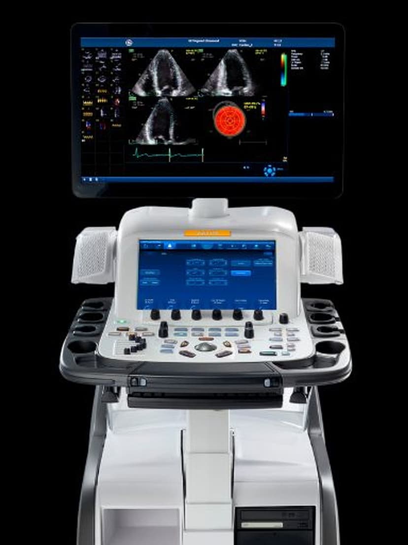
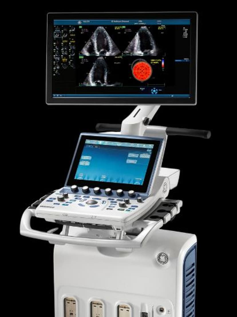
Compatible Systems
Specifications
Wide bandwidth covers deep adult cardiac structures and high-resolution valve leaflet detail from the esophageal position.
Standard TEE sector angle captures four-chamber and long-axis views from mid-esophageal position.
Reaches descending aorta and posterior cardiac structures from the esophageal approach.
Full Doppler suite quantifies valvular gradients, regurgitation, and intracardiac flow.
D-Pin interface connects to GE Vivid cardiac ultrasound platforms.
Applications
Intraoperative Cardiac Monitoring
The 10T-D provides real-time TEE imaging during cardiac surgery, valve repair, and structural heart interventions. The 90-degree sector view captures standard mid-esophageal views including four-chamber, two-chamber, and long-axis planes. Surgeons and anesthesiologists use these views to assess ventricular function, confirm valve competence after repair, and detect complications like air embolism before chest closure.
Valve Assessment
At 7–10 MHz, the 10T-D resolves mitral and aortic valve leaflet thickness, coaptation defects, and vegetations that transthoracic imaging may miss. Color Doppler maps regurgitant jets with higher spatial resolution than chest-wall approaches, and the posterior esophageal position eliminates rib and lung artifact. PW and CW Doppler quantify transvalvular gradients for stenosis severity grading.
Adult & Pediatric TEE
The 3.3 MHz low end provides sufficient penetration for adult patients with larger chest dimensions, while the compact probe shaft accommodates larger pediatric patients for congenital heart disease evaluation. Mid-esophageal and transgastric views characterize septal defects, intracardiac shunts, and great vessel anatomy. The D-Pin connector pairs the 10T-D exclusively with Vivid-series cardiac platforms.
Our Partners


Need this probe for your system?
Request a quote with pricing, compatibility verification, and delivery timeline.