
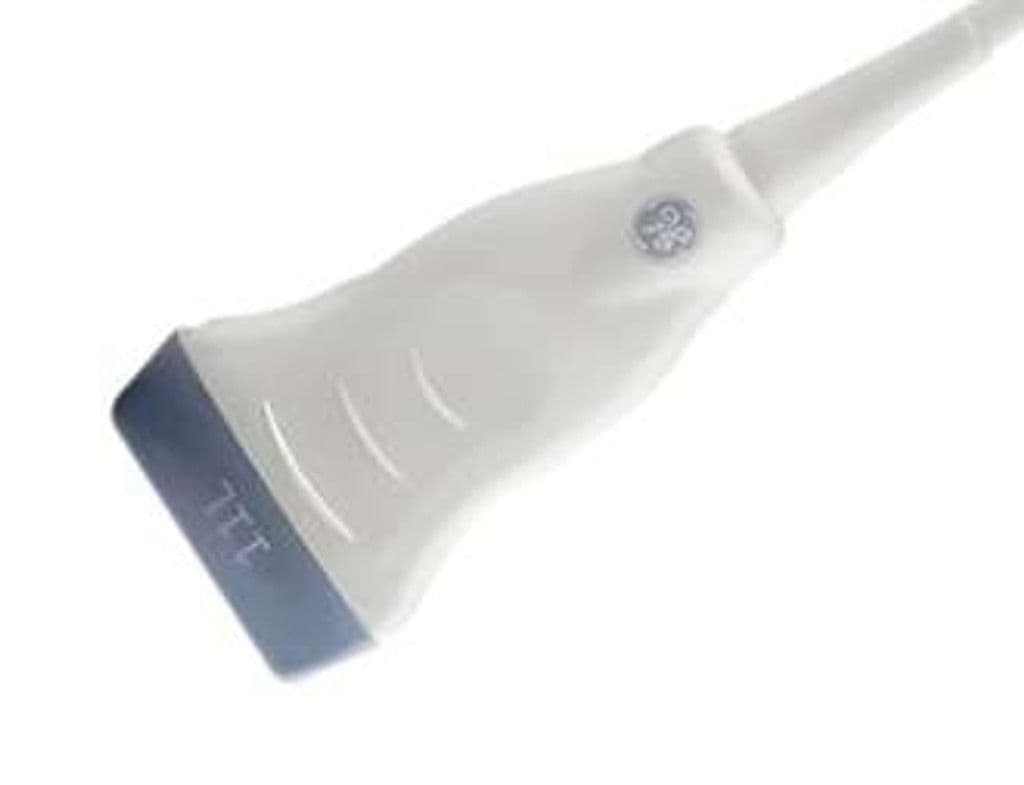
GE 11L-D
The 11L-D brings high-frequency linear imaging to GE's Vivid cardiac systems, adding vascular and small parts capability to platforms primarily known for echocardiography. The 4–12 MHz bandwidth handles carotid duplex, thyroid nodule characterization, and breast lesion evaluation without switching to a separate general imaging system. Harmonic imaging support reduces near-field clutter, and the slim probe head fits into tight scanning windows around the neck and extremities.
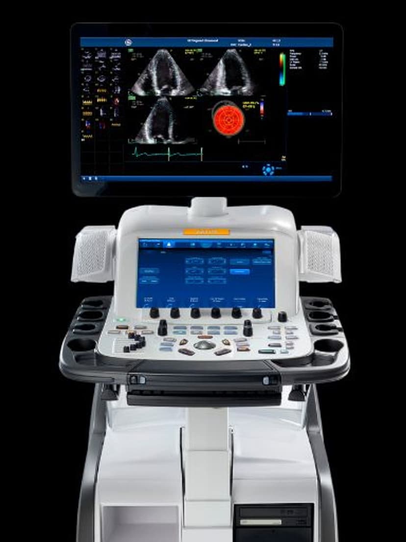
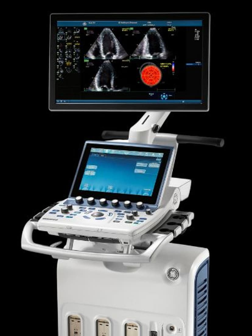
Compatible Systems
Specifications
Wideband range spans deep vascular imaging through high-resolution superficial tissue assessment.
Harmonic mode reduces near-field clutter for cleaner superficial tissue and breast imaging.
Sufficient depth for peripheral vascular work and deeper MSK structures.
D-Pin interface connects to GE Vivid E95 and Vivid S70 cardiac systems.
Applications
Vascular Imaging
Carotid duplex scanning on the Vivid platform allows cardiology practices to add vascular screening without a dedicated general imaging system. B-mode plaque characterization and spectral Doppler waveform analysis support stroke risk stratification. The 4 MHz low end reaches the deeper subclavian and femoral vessels for full peripheral vascular assessment.
Breast & Thyroid Small Parts
High-frequency operation at 10–12 MHz resolves thyroid nodules for TI-RADS scoring and breast lesions for BI-RADS classification. Harmonic imaging reduces clutter artifacts in superficial tissue, improving lesion border definition. The linear geometry provides consistent caliper placement for accurate measurement of nodule dimensions.
Musculoskeletal Assessment
Tendon and ligament visualization benefits from the 12 MHz near-field resolution, with enough bandwidth to image deeper structures like the supraspinatus in the shoulder. Dynamic scanning of the rotator cuff, Achilles tendon, and lateral epicondyle guides clinical decision-making for sports medicine and orthopedic referrals. Color Doppler identifies hyperemia in inflammatory conditions and post-surgical sites.
Our Partners


Need this probe for your system?
Request a quote with pricing, compatibility verification, and delivery timeline.