
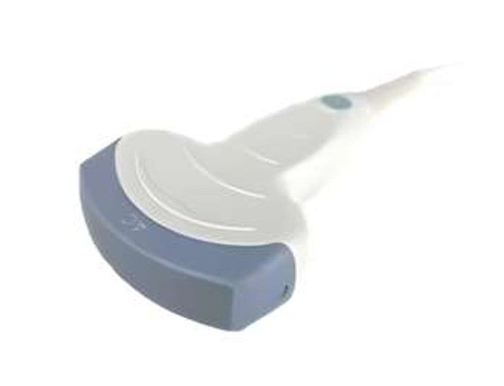
GE 4C-RS
The 4C-RS is GE's general-purpose convex transducer, pairing a 2.0 – 5.0 MHz bandwidth with a 58-degree sector angle and 21 x 24 mm footprint for broad anatomical coverage. Depth penetration reaches 30 cm, making it effective for large-habitus abdominal scans and second-/third-trimester obstetric evaluations. B-mode, Color Doppler, and PW Doppler modes support both morphological and hemodynamic assessment across abdominal and pelvic organs.
Specifications
Low-to-mid frequency range penetrates up to 30 cm for deep abdominal and obstetric imaging.
Wide sector angle captures large anatomical regions in a single sweep.
Moderate contact area balances acoustic coupling with intercostal access.
Full-depth penetration supports imaging of large-habitus patients.
Standard mode set for morphological and hemodynamic abdominal evaluation.
RS-Pin interface connects to the broadest range of GE general-imaging and OB/GYN systems.
Applications
Abdominal Imaging
The 58-degree field of view covers the full liver, kidneys, spleen, and retroperitoneum in standard scanning planes. At 1–5 MHz, the probe balances resolution for parenchymal detail with penetration through abdominal wall fat. Color Doppler evaluates hepatic and renal vasculature for portal hypertension screening and renal artery stenosis workup.
Obstetric & Gynecological Imaging
Second- and third-trimester fetal anatomy surveys benefit from the wide sector angle and 30 cm depth. Biometric measurements (BPD, HC, AC, FL) are performed in B-mode, while umbilical artery Doppler assesses placental perfusion. Gynecological applications include uterine morphology, fibroid mapping, and adnexal mass characterization.
Urological Imaging
Provides transabdominal views of the bladder, kidneys, and prostate gland. Pre- and post-void volume measurements guide management of urinary retention. Color Doppler detects renal vascular abnormalities, and B-mode identifies hydronephrosis, calculi, and bladder wall thickening.
Our Partners


Need this probe for your system?
Request a quote with pricing, compatibility verification, and delivery timeline.