
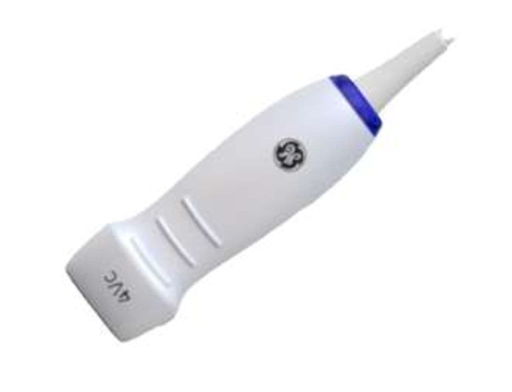
GE 4V-D
The 4V-D is a volume phased array that acquires full cardiac datasets in real time, enabling 4D visualization of chamber geometry, wall motion, and valvular function that 2D imaging cannot capture in a single acquisition. The 21 x 24 mm footprint fits standard intercostal windows, and the 1.5–5 MHz bandwidth covers both adult and larger pediatric patients. Volume acquisition supports automated quantification workflows like 4D Auto LVQ on the Vivid E95, reducing exam time for ejection fraction, volumes, and strain analysis.
Compatible Systems
Specifications
Low-frequency penetration reaches all cardiac structures in adult patients; higher frequencies resolve valve and endocardial detail.
Standard cardiac sector angle captures four-chamber and long-axis views from the apical window.
Deep penetration images the entire heart in larger adult patients and reaches the descending aorta.
Compact face fits standard intercostal spaces for apical, parasternal, and subcostal windows.
Full 4D volume mode enables real-time three-dimensional cardiac imaging and automated LV quantification.
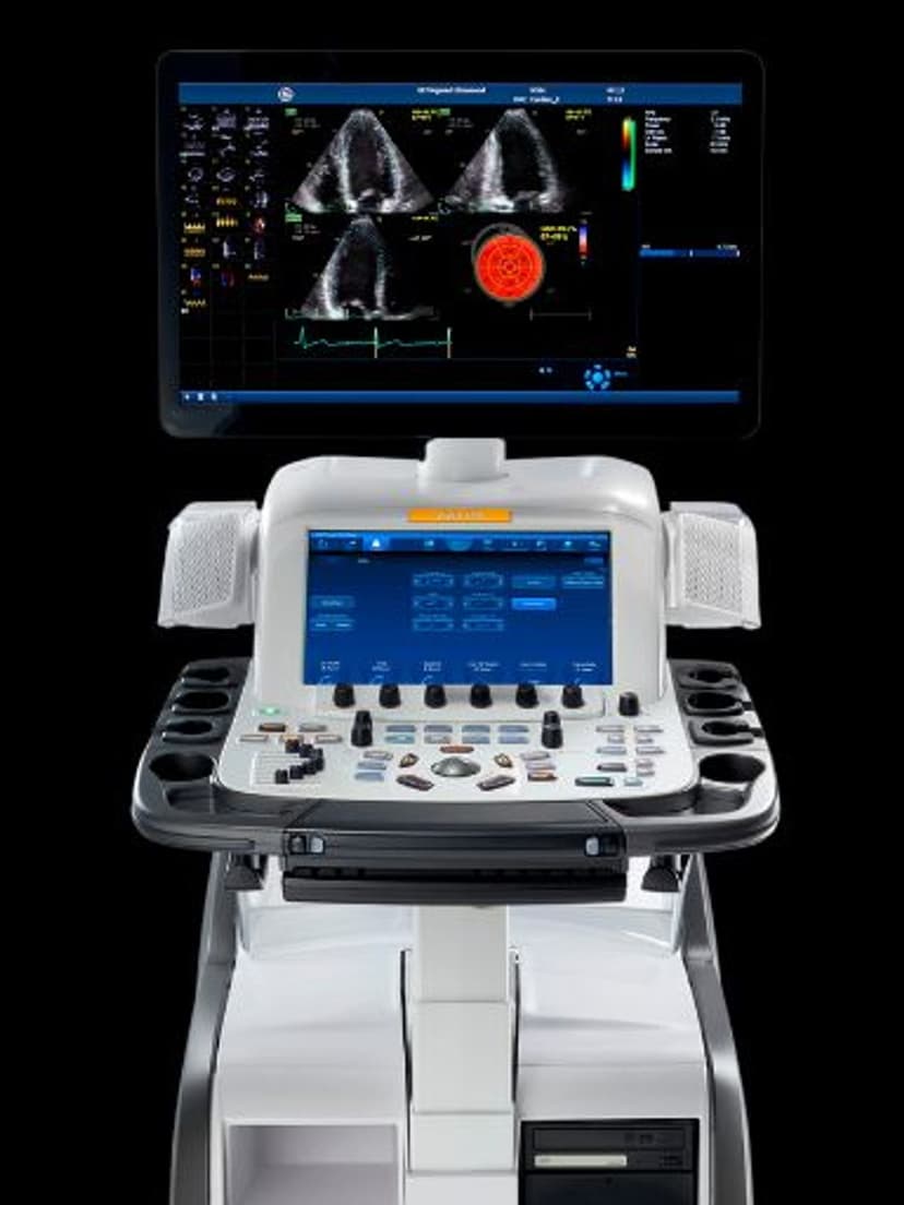
D-Pin interface connects to GE Vivid cardiac platforms including the E95.
Applications
4D Transthoracic Echocardiography
Real-time volume acquisition captures the full left ventricle in a single heartbeat, eliminating foreshortening and geometric assumptions inherent in 2D biplane methods. The 4V-D supports 4D Auto LVQ for automated ejection fraction, end-diastolic volume, and end-systolic volume measurements. Clinicians can crop and rotate the dataset post-acquisition to extract any standard view plane without rescanning.
LVO Contrast Imaging
Optimized for left ventricular opacification with ultrasound contrast agents, the 4V-D's low mechanical index settings preserve microbubble integrity while the volume dataset captures endocardial borders from all angles simultaneously. This improves wall motion scoring in patients with difficult acoustic windows where native 2D imaging produces suboptimal endocardial definition.
Stress Echocardiography
Volume acquisition at each stage of a stress protocol, whether pharmacologic or exercise, captures all 17 myocardial segments in fewer acquisitions than standard 2D stress echo. The 90-degree sector angle and 30 cm depth accommodate the full heart even during tachycardic stress states. Side-by-side 4D comparison of rest, peak, and recovery stages improves wall motion abnormality detection.
Our Partners


Need this probe for your system?
Request a quote with pricing, compatibility verification, and delivery timeline.