
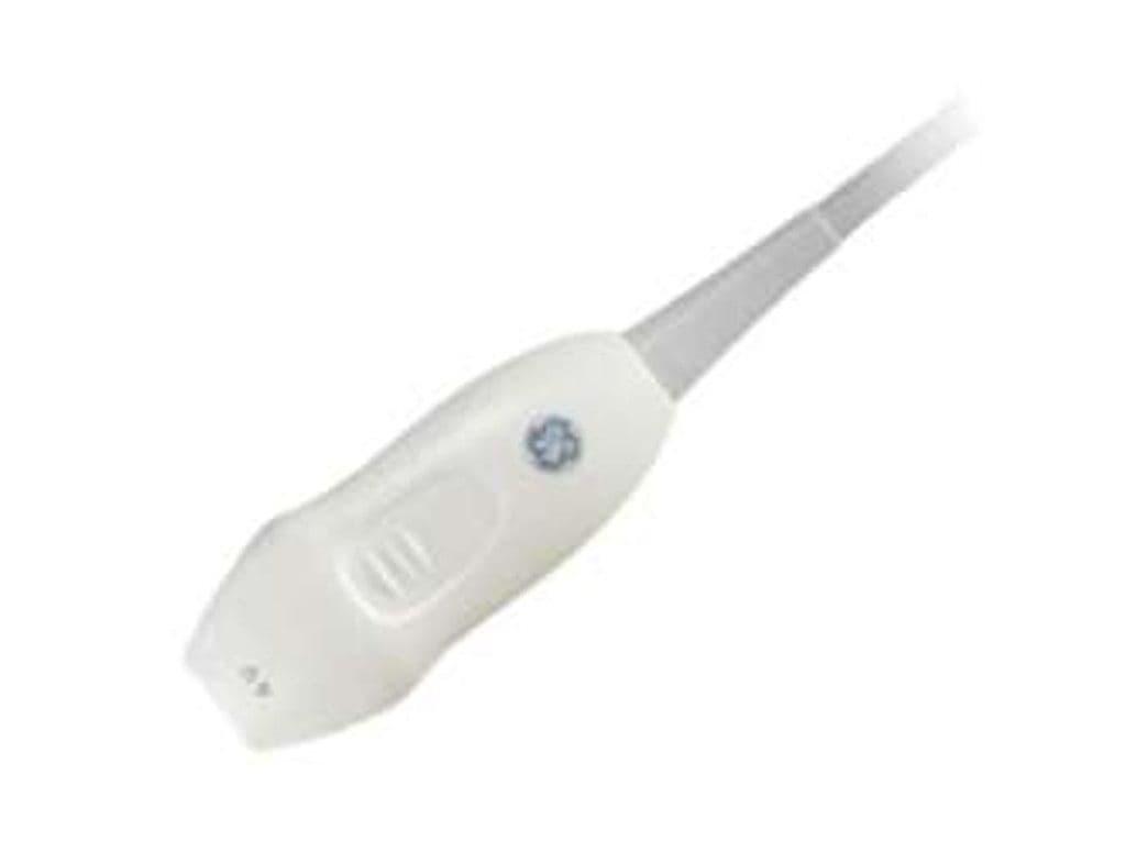
GE 4Vc-D
The 4Vc-D brings real-time 4D volume imaging to the Vivid platform through a compact phased array element with XDclear crystal technology. The 1.4 – 5.2 MHz bandwidth and 90-degree field of view cover standard TTE windows including parasternal, apical, and subcostal approaches. The small probe head accesses narrow intercostal spaces in pediatric and thin adult patients, while 30 cm depth penetration handles larger body habitus without sacrificing frame rate.
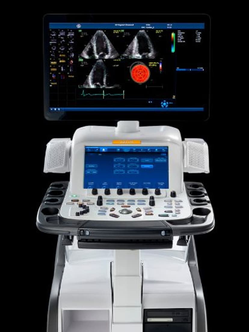
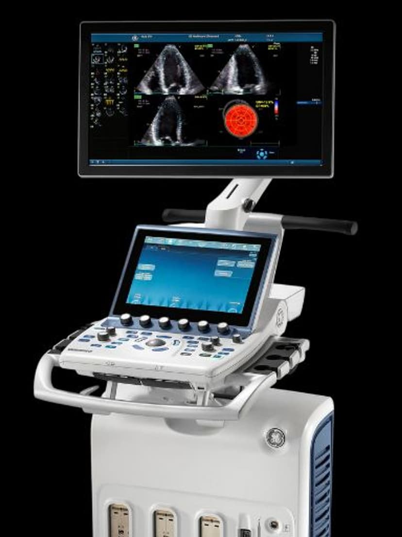
Compatible Systems
Specifications
Low-frequency range penetrates through the chest wall for adult and pediatric cardiac imaging.
Wide sector covers the full cardiac anatomy in standard transthoracic windows.
4D volume mode adds real-time 3D cardiac visualization to standard 2D and Doppler protocols.
Full-depth penetration accommodates large-habitus adult echocardiography.
Single-crystal technology improves sensitivity and bandwidth for higher frame rates during volume acquisition.
D-Pin interface connects to GE Vivid-series cardiac systems.
Applications
Adult Echocardiography
Real-time 4D volume acquisition captures full-volume datasets of the left ventricle for automated EF calculation and wall motion analysis. Standard 2D and M-mode imaging covers parasternal long-axis, short-axis, apical four-chamber, and subcostal views. Color and PW Doppler quantify valvular regurgitation severity and transmitral flow patterns for diastolic function assessment.
Pediatric Cardiac Imaging
The compact probe head fits between narrow pediatric rib spaces where adult-sized phased arrays cannot achieve acoustic contact. Higher-frequency operation at 6.0 MHz resolves small cardiac structures including the atrial septum, VSD margins, and valve leaflets in neonatal and pediatric patients. 4D imaging aids in spatial orientation of congenital heart defects.
4D Volume Echocardiography
Full-volume 4D datasets enable offline multiplanar reconstruction for mitral valve analysis, left atrial appendage assessment, and surgical planning. Real-time 3D rendering visualizes valve morphology from multiple perspectives without transducer repositioning. Volume-rendered datasets support strain analysis and synchrony evaluation on Vivid-series systems with EchoPAC.
Our Partners


Need this probe for your system?
Request a quote with pricing, compatibility verification, and delivery timeline.