
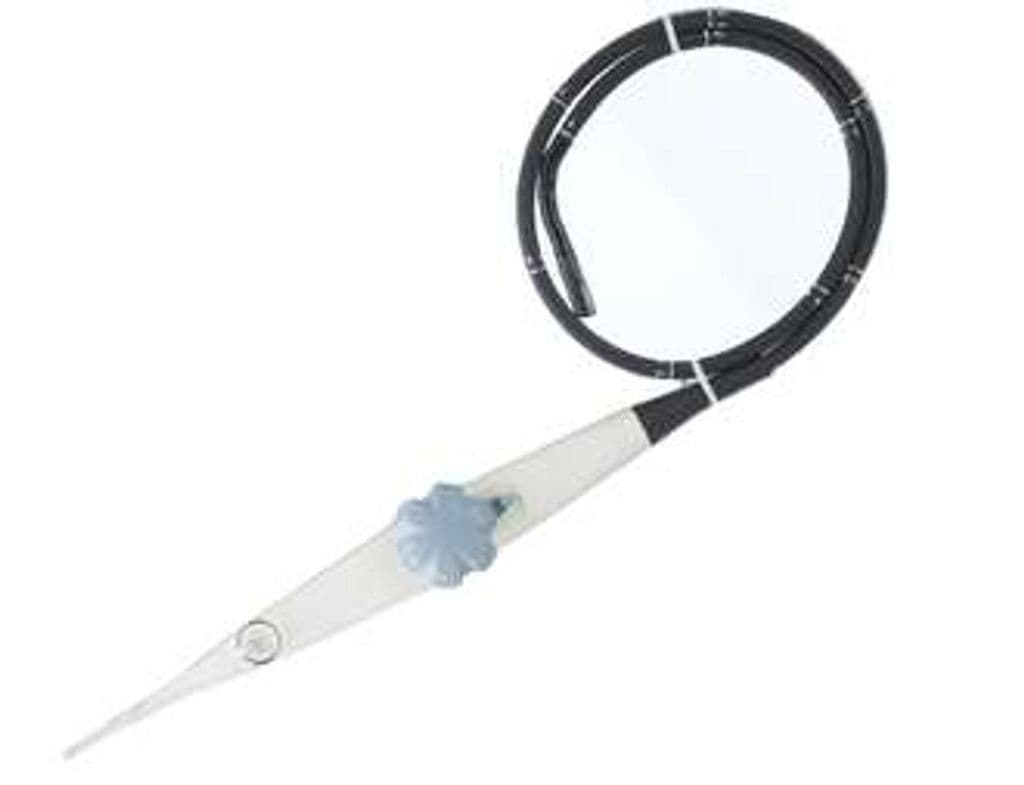
GE 6VT-D
The 6VT-D is GE's electronic 4D multiplane TEE probe, using an active matrix array to acquire full-volume cardiac datasets in real time without mechanical rotation. The 3–8 MHz frequency range and 90-degree field of view cover standard mid-esophageal and transgastric imaging planes for structural heart assessment. Real-time 4D rendering of valve anatomy, septal defects, and left atrial appendage morphology supports both diagnostic echocardiography and intraprocedural guidance for structural heart interventions. D-Pin connectivity restricts it to the Vivid E95, S70, and IQ platforms.
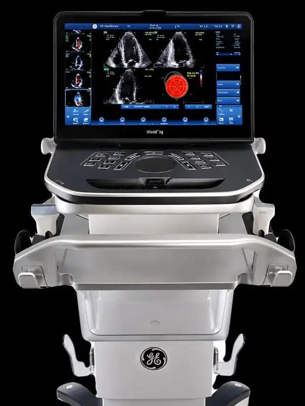
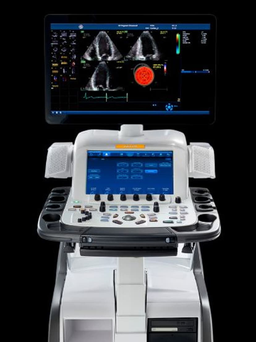
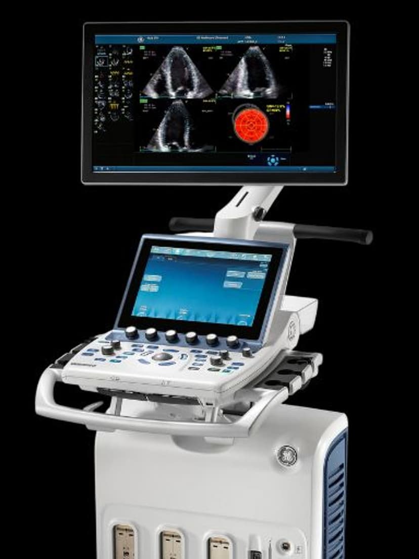
Compatible Systems
Specifications
Mid-frequency range balances penetration for transgastric views with resolution for valve leaflet detail.
Electronic volumetric acquisition without mechanical rotation for real-time 3D cardiac imaging.
Wide sector captures complete cardiac structures from standard TEE imaging windows.
Full cardiac TEE mode set including real-time volumetric rendering for structural assessment.
Sufficient depth for mid-esophageal and transgastric cardiac views.
D-Pin interface connects to GE Vivid E95, S70, and IQ cardiac platforms.
Applications
4D Transesophageal Echocardiography
The active matrix array acquires volumetric datasets that can be cropped and rotated post-acquisition to view valve anatomy from any angle. Real-time 4D visualization of mitral valve leaflet motion, commissural anatomy, and regurgitant orifice geometry informs surgical and transcatheter repair planning. The electronic multiplane capability eliminates mechanical rotation, allowing rapid switching between standard 2D imaging planes and full-volume 4D acquisitions.
Structural Heart Intervention Guidance
Intraprocedural TEE guidance during transcatheter aortic valve replacement, MitraClip deployment, and left atrial appendage closure relies on real-time 3D visualization of device positioning relative to cardiac anatomy. The 6VT-D provides the en-face views of the mitral and aortic valves that are essential for guiding device placement. Color Doppler overlay on 4D volumes maps paravalvular leak location and severity during and after deployment.
Left Ventricular Opacification & Contrast
Contrast-enhanced imaging with the 6VT-D improves endocardial border delineation for accurate ejection fraction measurement in patients with suboptimal acoustic windows. LVO protocols enhance detection of apical thrombus and wall motion abnormalities that may be missed on standard imaging. The 3–8 MHz range supports the low mechanical index required for microbubble persistence during contrast examinations.
Our Partners


Need this probe for your system?
Request a quote with pricing, compatibility verification, and delivery timeline.