
GE 9T
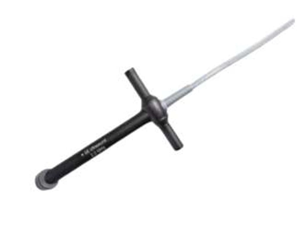
The 9T is GE's multiplane transesophageal echocardiography probe, housing a phased array element in a 7.5 x 10.7 mm tip with a 37.5 mm insertion length. The 3–10 MHz bandwidth provides near-field resolution for mitral and aortic valve morphology at higher frequencies and full 14 cm penetration at lower frequencies for left atrial appendage and descending aorta imaging. Multiplane rotation from 0 to 180 degrees captures standard TEE views without repositioning the probe shaft.
Compatible Systems
Specifications
Broad bandwidth covers near-field valve detail at 10 MHz and full-chamber penetration at 3 MHz.
Wide sector angle captures the full cardiac chamber in standard transesophageal windows.
Compact tip dimensions enable use in adult and larger pediatric patients.
Short insertion segment minimizes esophageal contact area during prolonged intraoperative monitoring.
Sufficient depth for left atrial appendage, descending aorta, and transgastric views.
Standard Doppler suite supports hemodynamic quantification during TEE procedures.
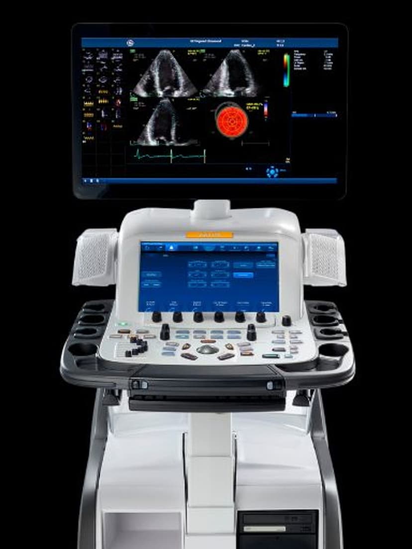
D-Pin interface connects to the GE Vivid E95 cardiac platform.
Applications
Transesophageal Echocardiography
Standard 20-view TEE protocol acquisition from the mid-esophageal, transgastric, and upper esophageal positions. Multiplane rotation sweeps through 0–180 degrees for aortic valve short-axis, mitral commissural, bicaval, and four-chamber views. The 3–10 MHz bandwidth allows operators to shift between high-resolution near-field imaging of valve leaflets and deeper penetration for left atrial appendage thrombus screening.
Intraoperative Cardiac Monitoring
Real-time TEE during cardiac surgery guides valve repair, assesses de-airing after cardiopulmonary bypass, and confirms graft anastomosis patency. Immediate post-repair imaging identifies residual regurgitation or paravalvular leak before chest closure. Color Doppler and PW Doppler quantify flow across repaired or replaced valves in the operating room.
Structural Heart Intervention Guidance
TEE provides real-time imaging during percutaneous procedures such as transcatheter aortic valve replacement (TAVR), MitraClip deployment, and atrial septal defect closure. The 90-degree field of view and multiplane capability localize guide wires, catheters, and delivery devices relative to cardiac anatomy. High-frequency imaging resolves device positioning against the interatrial septum and valve annulus.
Our Partners


Need this probe for your system?
Request a quote with pricing, compatibility verification, and delivery timeline.