
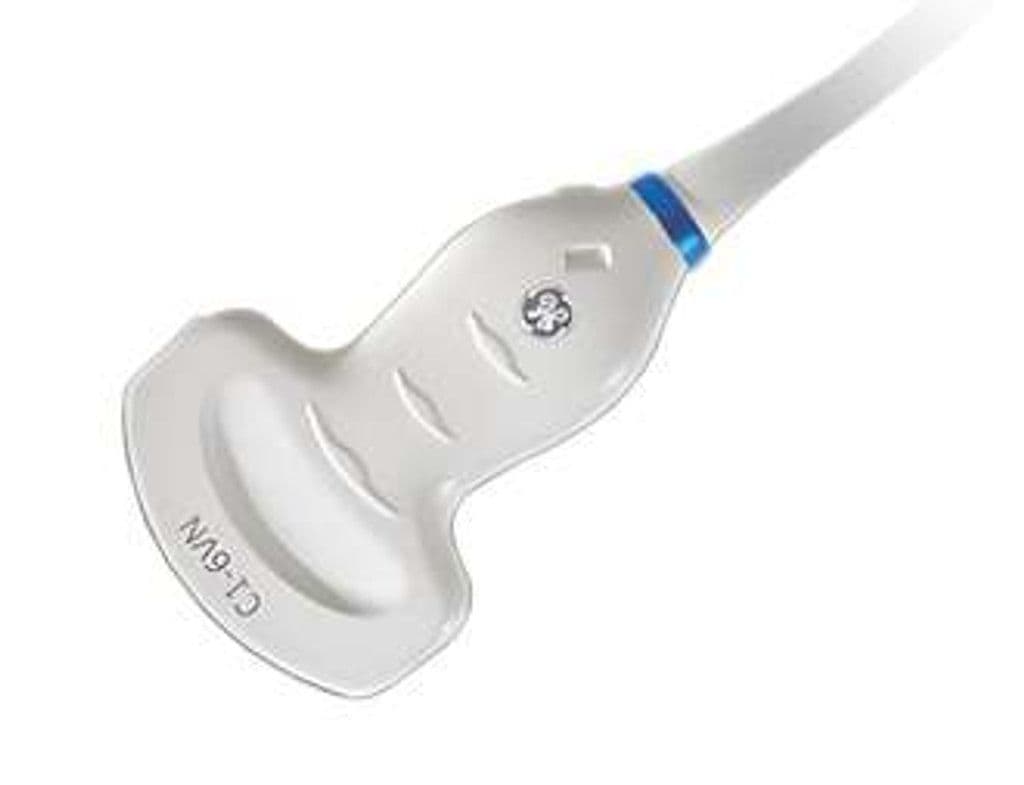
GE C1-6VN-D
The C1-6VN-D pairs XDclear transducer technology with a volume-capable curved array for high-resolution 3D/4D imaging on GE D-pin platforms. XDclear construction uses single-crystal materials and advanced acoustic matching layers for wider bandwidth and improved sensitivity compared to standard piezoelectric elements. The 30 cm depth capability and 70-degree field of view support deep abdominal imaging, while volume acquisition enables HDlive rendering and STIC fetal cardiac analysis.
Compatible Systems


Specifications
Wideband XDclear elements provide improved sensitivity for deep abdominal and obstetric imaging.
Wide field captures full organ cross-sections and fetal anatomy in a single sweep.
Extended depth supports imaging in bariatric patients and deep retroperitoneal structures.
Full imaging suite includes volumetric acquisition for HDlive rendering and STIC cardiac analysis.
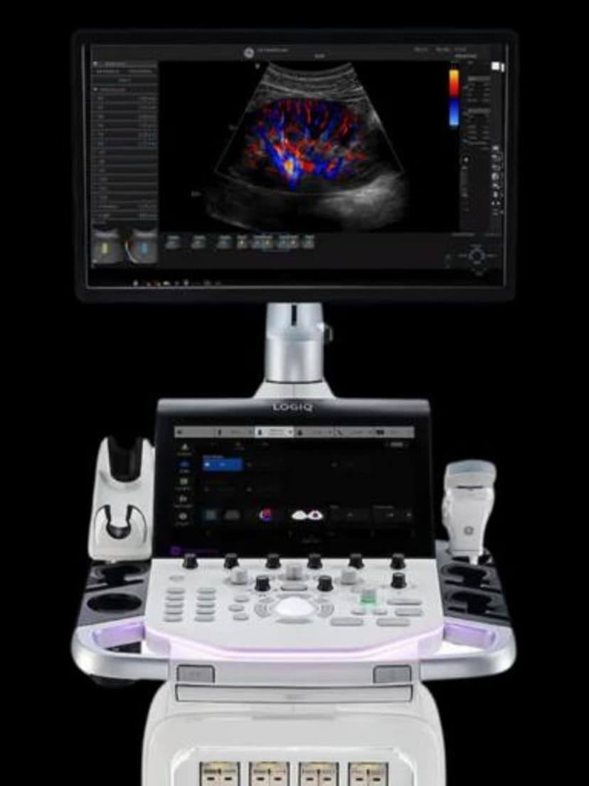
D-pin interface connects to GE LOGIQ, Vivid, and Voluson D-pin systems with volume rendering capability.
Applications
Obstetric 3D/4D Volume Imaging
XDclear element technology delivers improved signal-to-noise for HDlive and HDlive Silhouette rendering, producing photorealistic fetal surface images and skeletal visualization. STIC volume acquisition captures a complete fetal cardiac cycle for offline four-chamber and outflow tract review.
Abdominal and Urological Imaging
The 30 cm maximum depth accommodates bariatric patients and deep retroperitoneal anatomy. Standard abdominal protocols for liver, kidney, gallbladder, and pancreas benefit from XDclear bandwidth. Volume mode adds coronal and oblique plane reconstruction for complex mass characterization.
Gynecological Assessment
Transabdominal 3D volume acquisition captures uterine morphology for Mullerian anomaly classification, fibroid mapping, and IUD localization. The C1-6VN-D's wider bandwidth compared to standard convex probes improves tissue differentiation in pelvic imaging.
Our Partners


Need this probe for your system?
Request a quote with pricing, compatibility verification, and delivery timeline.