
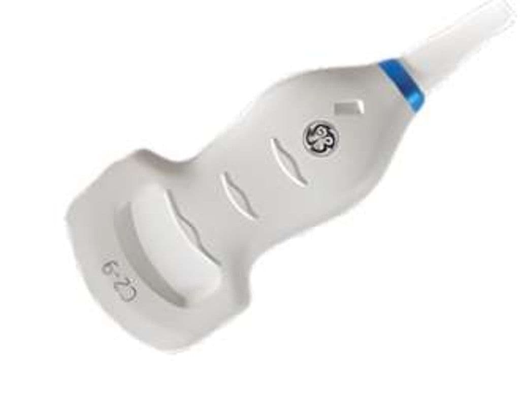
GE C2-9-D
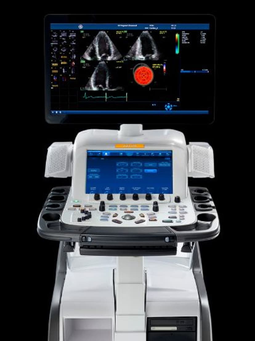
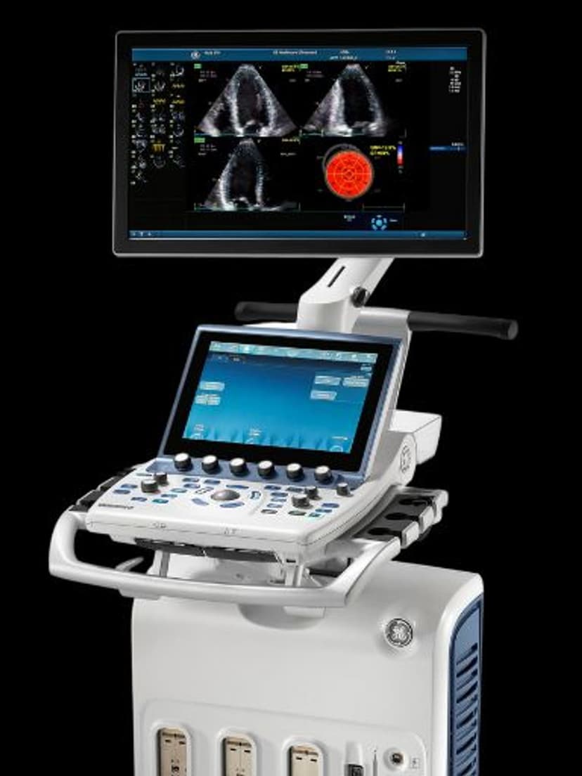
The C2-9-D delivers 2.0–9.0 MHz bandwidth through a compact curved array with a 70-degree sector and 20 cm depth range. The wideband design covers deep liver parenchyma at 2.0 MHz through near-field fetal anatomy at 9.0 MHz without switching transducers. Compatible with the Vivid E95, Vivid S70, LOGIQ E10, and LOGIQ Fortis, the C2-9-D serves as the primary abdominal and obstetric probe on GE's flagship D-Pin platforms.


Specifications
Wideband range covers deep abdominal penetration through near-field obstetric detail in one probe.
Wide sector captures complete organ cross-sections and fetal anatomy in single image frames.
Deep penetration accommodates all body habitus for abdominal and obstetric imaging.
Full diagnostic mode set for abdominal morphology, obstetric biometry, and vascular hemodynamics.
D-Pin interface connects to GE flagship Vivid and LOGIQ systems.
Applications
Abdominal Imaging
The 2.0 MHz low end penetrates to 20 cm for liver, kidney, spleen, and retroperitoneal assessment in all patient body types. At 6–9 MHz, superficial structures including the gallbladder wall, renal cortex, and bowel loops image with detail sufficient for grading fatty liver and detecting small calculi. The 70-degree sector captures complete organ cross-sections in single image frames for efficient abdominal surveys.
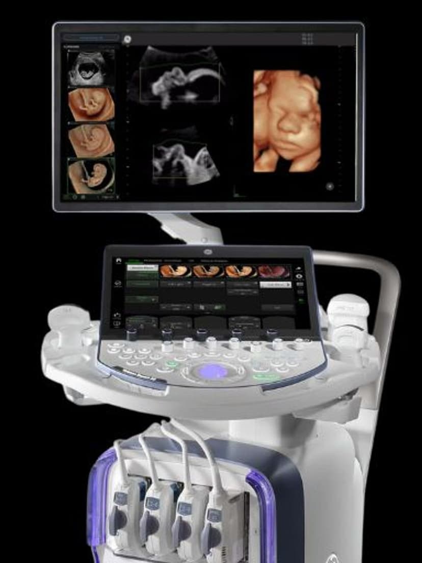
OB/GYN Imaging
Obstetric scanning across all trimesters uses the full bandwidth: 6–9 MHz for first-trimester dating and nuchal translucency, 3–5 MHz for second- and third-trimester anatomy surveys and biometric measurements. The curved array geometry follows the uterine contour for consistent contact during fetal growth assessment. Color Doppler evaluates umbilical artery, middle cerebral artery, and uterine artery flow for fetal well-being monitoring.
Abdominal Vascular Assessment
PW Doppler measures portal vein velocity, hepatic vein waveforms, and renal artery resistive indices for evaluating portal hypertension, hepatic congestion, and renal artery stenosis. The 2.0 MHz low end reaches the aorta and iliac bifurcation in larger patients for aneurysm screening. Color Doppler maps flow direction and detects thrombosis in the mesenteric, splenic, and renal vasculature.
Our Partners


Need this probe for your system?
Request a quote with pricing, compatibility verification, and delivery timeline.