
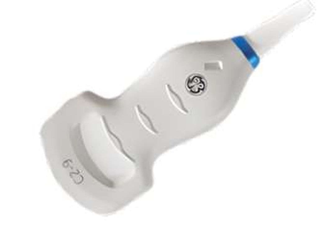
GE C2-9VN-D
The C2-9VN-D adds mechanical volume sweep capability to a curved array, enabling 3D/4D rendering of fetal anatomy, abdominal organs, and pelvic structures. The 2.0 – 9.0 MHz bandwidth covers deep abdominal scans at the low end and higher-resolution superficial OB/GYN imaging at the upper range. The 70-degree 2D field of view expands into a full volume dataset for HDlive rendering, multiplanar reconstruction, and volumetric organ measurements on compatible Voluson and LOGIQ systems.
Compatible Systems


Specifications
Wide bandwidth covers deep abdominal work and higher-resolution obstetric imaging in one volume probe.
Standard curved array sector angle captures full organ views for abdominal and pelvic exams.
Sufficient penetration for adult abdominal organs, retroperitoneal structures, and larger maternal habitus.
Volume modes enable HDlive rendering, multiplanar reconstruction, and automated organ volumetrics.
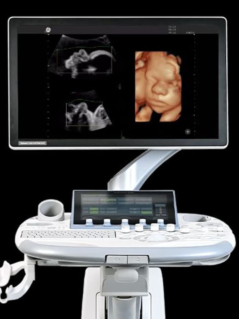
D-Pin interface connects to GE's Voluson and LOGIQ flagship platforms for 3D/4D imaging.
Applications
Obstetric 3D/4D Imaging
Volume acquisition captures the full fetal face, spine, and extremities for 3D surface rendering and multiplanar analysis. HDlive-compatible datasets produce realistic fetal images that improve parent communication and aid in detecting facial clefts and skeletal anomalies. The 2.3 MHz low end penetrates through larger maternal habitus, while 8.4 MHz resolves fine fetal structures in early second trimester.
Gynecologic Pelvic Imaging
3D volume rendering reconstructs the uterine cavity in coronal planes that are inaccessible with 2D transabdominal scanning alone. This coronal view differentiates septate from bicornuate uterine anomalies and maps the location of submucosal fibroids relative to the endometrial cavity. The wide bandwidth supports both transabdominal and low-frequency transvaginal approaches depending on clinical need.
Abdominal & Urological Imaging
In 2D mode, the C2-9VN-D functions as a standard curved array for liver, kidney, and bladder assessment. The 70-degree sector covers the full right hepatic lobe, and 2.3 MHz penetration reaches deep retroperitoneal structures. Volume mode adds renal volumetric measurements and 3D bladder volume calculations that reduce the need for catheterization-based residual volume testing.
Our Partners


Need this probe for your system?
Request a quote with pricing, compatibility verification, and delivery timeline.