
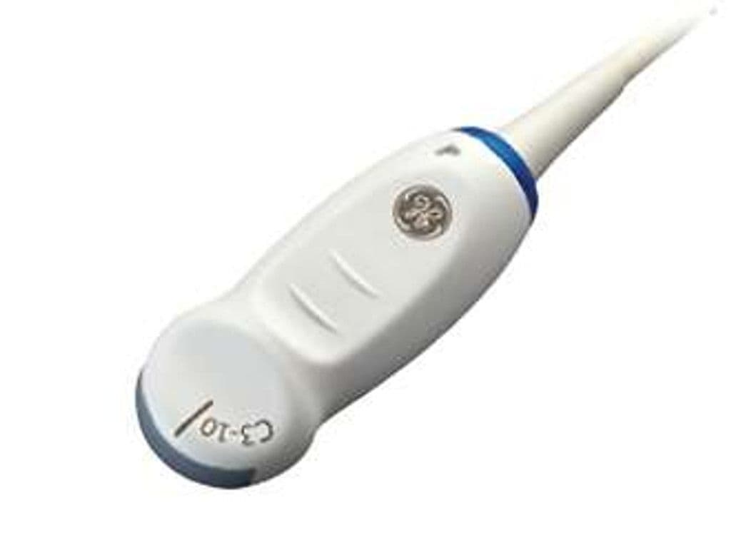
GE C3-10-D
The C3-10-D is a compact curved array transducer with a 3.0–10.0 MHz bandwidth tailored for pediatric and neonatal imaging. Its small footprint and 50-degree field of view suit neonatal cranial access through the fontanelle and pediatric abdominal scans where a standard convex probe is too large. The high upper frequency resolves superficial structures like the neonatal brain parenchyma, while 3.0 MHz maintains adequate penetration for older pediatric abdominal exams at depths up to 12 cm.


Specifications
Extended bandwidth covers neonatal superficial imaging through deeper pediatric abdominal penetration.
Compact sector angle sized for neonatal fontanelle access and small pediatric anatomy.
Covers neonatal through school-age pediatric abdominal depths.
Doppler suite supports vascular assessment and hemodynamic evaluation in pediatric patients.
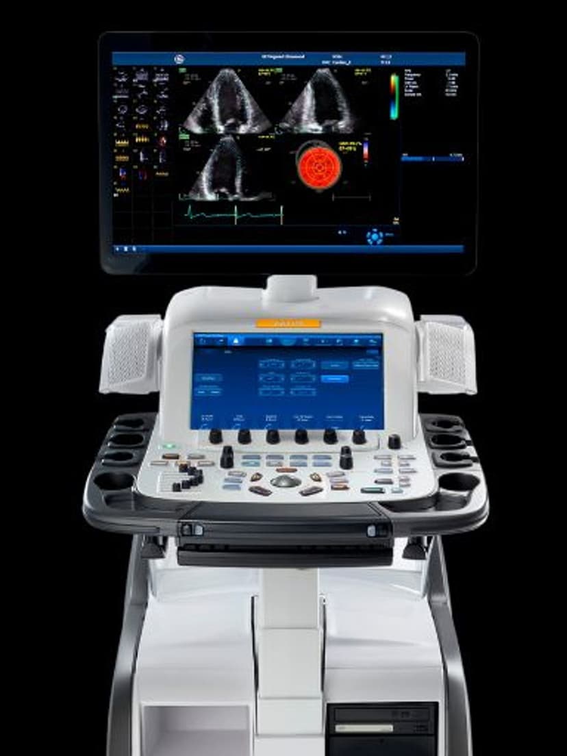
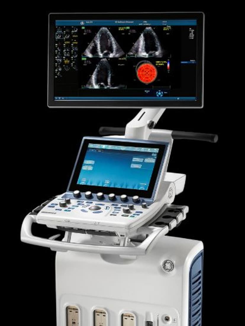
D-Pin connector for GE Vivid and LOGIQ premium platform systems.
Applications
Neonatal Cranial Imaging
The C3-10-D fits the anterior fontanelle for coronal and sagittal brain imaging in neonates. At 10.0 MHz, it resolves periventricular white matter, the germinal matrix, and ventricular margins with the detail needed to grade intraventricular hemorrhage. The curved array geometry provides a wider near-field view than linear probes, capturing both lateral ventricles in a single coronal sweep.
Pediatric Abdominal Imaging
For pediatric patients, the C3-10-D provides abdominal organ assessment including liver, kidneys, spleen, and appendix visualization. The 12 cm depth range covers neonates through school-age children, and the compact footprint maneuvers between ribs and across small abdominal walls more easily than adult-sized convex probes. Color Doppler maps renal and hepatic vascularity.
Musculoskeletal and Vascular
The high-frequency range supports superficial MSK imaging of pediatric joints, tendons, and soft tissue masses. In vascular applications, the curved geometry provides a wider field than linear probes for mapping vessel courses, while the high bandwidth resolves vessel wall detail and supports accurate Doppler velocity measurements.
Our Partners


Need this probe for your system?
Request a quote with pricing, compatibility verification, and delivery timeline.