
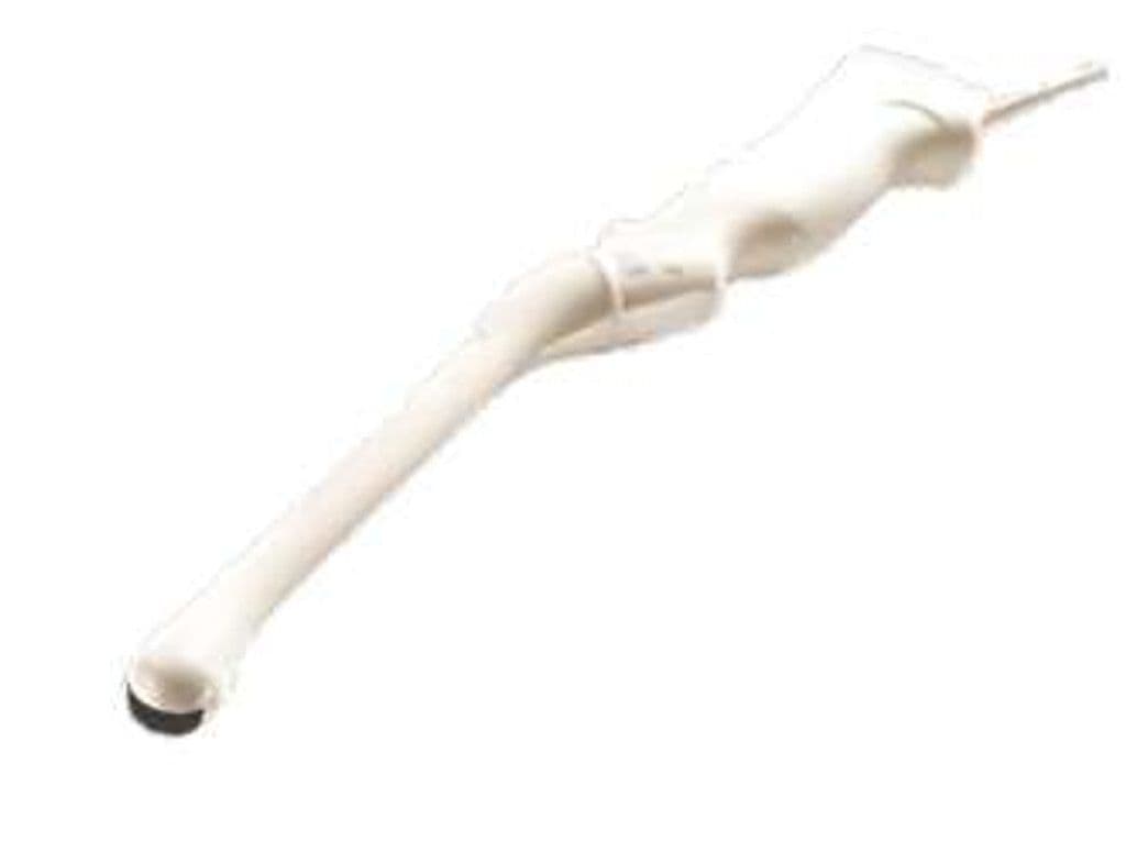
GE E8C-RS
The E8C-RS uses a microconvex array with a compact 16.9 x 21.2 mm footprint designed for transvaginal and transrectal approaches. The 128-degree sector angle captures the full uterus, ovaries, and adnexal regions without excessive probe manipulation. At 4.2 – 10.0 MHz, the bandwidth balances high-resolution endometrial and follicular imaging with enough penetration to assess deeper pelvic structures like the broad ligament and pelvic sidewall nodes.

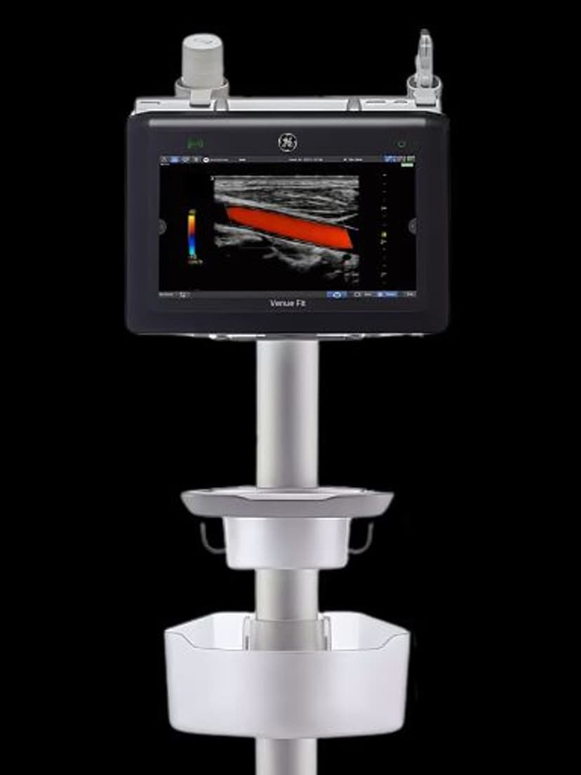
Specifications
Wide bandwidth covers deep pelvic masses and high-resolution endometrial detail from the endocavity approach.
Wide sector captures uterus, both ovaries, and adnexa with minimal probe rotation.
Compact microconvex tip improves patient comfort during transvaginal and transrectal exams.
Reaches deeper pelvic masses and broad ligament structures beyond the immediate endocavity field.
Doppler modes quantify ovarian and uterine artery flow for fertility and ectopic pregnancy assessment.
RS-Pin interface works across GE's broadest range of general imaging and OB/GYN platforms.
Applications
Transvaginal OB/GYN Imaging
The 128-degree field of view captures the uterus, both ovaries, and cul-de-sac in a single sweep. At 7–10 MHz, endometrial thickness measurements and follicular monitoring resolve with the detail needed for infertility workups and IVF cycle management. The 3.0 MHz low end reaches ectopic pregnancy locations and deeper pelvic masses that narrower-band endocavity probes may miss.
Early Pregnancy Assessment
High-frequency operation detects gestational sacs, yolk sacs, and fetal cardiac activity earlier than transabdominal approaches. Crown-rump length measurements at 6–12 weeks benefit from the short focal distance and high axial resolution of the endocavity approach. Doppler interrogation confirms corpus luteum vascularity and detects ectopic implantation sites.
Urological & Pelvic Floor Imaging
Transrectal positioning images the prostate, seminal vesicles, and bladder base. The compact 16.9 x 21.2 mm tip reduces patient discomfort compared to larger endocavity probes. Pelvic floor assessment during rest and Valsalva maneuvers evaluates organ descent and levator ani integrity for pre-surgical planning.
Our Partners


Need this probe for your system?
Request a quote with pricing, compatibility verification, and delivery timeline.